Stem Cell Therapy – When to Expect Results

Stem Cell Therapy – When to Expect Results

Stem cell therapy is one of the most promising areas of modern regenerative medicine. However, patients and even some physicians sometimes expect results too quickly, or conversely, anticipate unjustifiably delayed outcomes. To understand realistic timelines, it is important to consider the type of stem cells, their mechanism of action, the patient’s condition, and the nature of the disease.

How Stem Cells Work: Key Mechanisms

The result of therapy depends on what exactly the stem cells do after administration:

  • Direct regeneration – differentiation of administered cells into cells of the lost tissue (less common, slower).
  • Paracrine effect – secretion of growth factors, cytokines, and exosomes that stimulate the body’s own cells to recover (more common, faster).
  • Immunomodulation – suppression of pathological inflammation and normalization of immune response, creating conditions for regeneration.

Exosomes stem cells fcell-09-636136-g001

Factors Influencing the Time to See Results

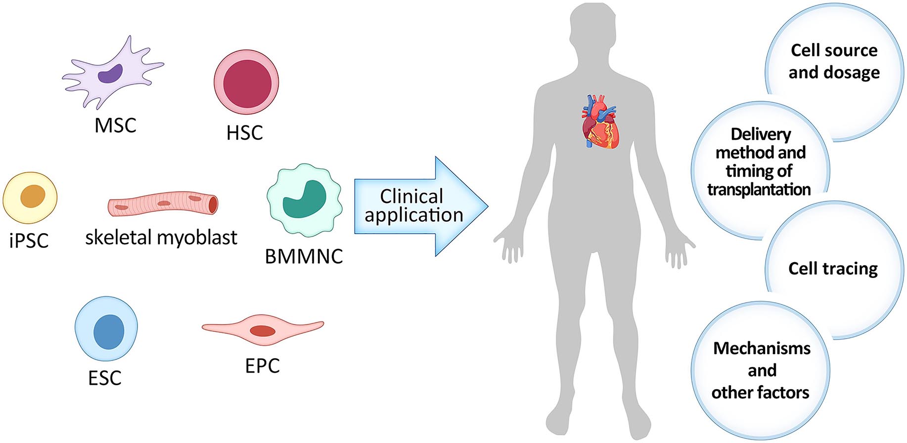
Type of Cells and Their Source

  • Mesenchymal stromal cells (MSC) – effects often start with anti-inflammatory changes (2–6 weeks), with a regenerative peak at 3–6 months.
  • Hematopoietic stem cells (HSC) – after transplantation, restoration of hematopoietic function takes 2–4 weeks, and immune stabilization takes months.
  • Induced pluripotent stem cells (iPSC) – timing depends on the stage of differentiation; often delayed for months, averaging from 3 months.
  • Exosomes and other extracellular vesicles – their action is often associated with triggering regeneration cascades, modulating inflammation, and improving cellular metabolism. Initial effects (reduced inflammation, improved skin condition, partial increase in energy) may be noticeable within 1–4 weeks, but structural changes (tissue regeneration, functional recovery) usually manifest within 1–3 months.
  • Narrowly differentiated cells (e.g., chondrocytes, neuroblasts, myoblasts) – may integrate more slowly. Engraftment and functionality take time; first improvements are possible after 3 months, with a peak effect at 6–12 months.
  • Mitochondrial therapy (transplantation of isolated mitochondria or stimulation of biogenesis) can produce a faster metabolic effect – sometimes within days or weeks, but a lasting functional result takes months.

Disease and Its Stage

  • Acute injury (e.g., limb ischemia) – clinical effect may appear within days or weeks.
  • Chronic degenerative diseases (osteoarthritis, neurodegeneration) – results develop slowly, usually over 3–12 months.
  • Autoimmune diseases – improvement may occur after immune stabilization, which takes 3–4 months.

Individual Factors

  • Age of the patient (younger → faster regeneration).
  • Immune status.
  • Comorbidities and use of medications (especially immunosuppressants or corticosteroids).

Exosomes stem cells how-long-does-stem-cell-therapy-last-for-knees-2

Typical Recovery Dynamics

Period After Therapy Biological Processes Possible Clinical Changes
0–2 weeks Immunomodulation, reduced inflammation, cell adaptation Reduction in pain, swelling, general improvement in well-being in some patients
1–3 months Paracrine stimulation of regeneration, activation of the body’s own progenitor cells Improved organ/tissue function, symptom reduction
3–6 months Maturation of new cells, restoration of microcirculation, tissue remodeling Maximum functional gain, stabilization of condition
6–12 months Long-term effects and maintenance of restored functions Consolidation of results, sometimes further improvement

Why There Is Not Always a Quick Result

  • Prolonged phase of cell integration and maturation.
  • Presence of irreversible tissue changes.
  • Strong chronic inflammation or active autoimmune process.
  • Insufficient cell dose or viability.

Stem Cell Path: From Administration to Clinical Effect

  • 0–24 hours — adaptation and survival of cells

What happens: cells encounter a new environment; some die, some attach to tissues.
What may slow it down: low cell viability, high inflammation, poor blood supply.
How to accelerate: use fresh/high-quality cells, reduce systemic inflammation beforehand, improve microcirculation.

  • 1–14 days — interaction with tissues and immunomodulation

What happens: cells secrete anti-inflammatory cytokines, begin paracrine effects on local cells.
What may slow it down: excessive immune response, autoimmune aggression.
How to accelerate: mild immunosuppression as indicated, use of exosomes or growth factors in parallel.

  • 2–8 weeks — stimulation of regeneration and activation of own progenitor cells

What happens: new vessels form, local stem cells are activated, inflammation decreases.
What may slow it down: fibrosis, chronic infections, vitamin/mineral deficiencies.
How to accelerate: physiotherapy, correction of deficiencies (D, B12, magnesium), PRP or growth factors.

  • 3–6 months — maturation and integration of new cells

What happens: administered and activated cells become mature tissue structures, functions are restored.
What may slow it down: tissue ischemia, recurrent inflammation, lack of stimulation (physical inactivity).
How to accelerate: physical therapy, rehabilitation, supportive therapy (angioprotectors, mitochondrial stimulators).

  • 6–12 months — effect stabilization

What happens: consolidation of functional improvements, tissue adaptation to new conditions.
What may slow it down: disease recurrence, unaddressed risk factors.
How to accelerate/maintain: repeat therapy courses (if indicated), control of chronic diseases, healthy lifestyle.

Exosomes stem cells 12015_2020_10110_Fig4_HTML

Survival of Biobank-Derived Products in the Body


The survival and “behavior” of administered bioproducts depend greatly on the product type, delivery method, and condition of the recipient tissue. Below is a detailed yet practical breakdown by type: expected lifespan in the body, how quickly their quantity and functionality usually decline, and what factors influence these parameters.
Hematopoietic stem cells (HSC) — when transplantation is performed correctly, they engraft and persist for a lifetime.
Mesenchymal stromal cells (MSC) — after intravenous infusions, most either die or “settle” in the lungs within 24–72 hours, though their paracrine effect can last for weeks to months; with local delivery, some cells may survive for weeks to months, but the proportion of long-term integrated cells is usually small.
• iPSC derivatives — survival time varies: with successful integration (e.g., neurons, cardiomyocytes), cells can survive for months to years, but this depends on immunity and product safety.
• Narrowly differentiated cells (tissue-specific progenitors) — with proper delivery and immunosuppression, they may integrate and function for extended periods (months to years).
• Exosomes / extracellular vesicles (EVs) — in the bloodstream, they have a short circulation time (minutes to hours) but can induce biological responses lasting days to weeks.

1. Mesenchymal Stromal Cells (MSC — bone marrow, adipose tissue, umbilical tissue)
What happens after administration:
• Intravenous (IV) delivery: most MSCs are trapped in the lung capillaries (“first-pass pulmonary trap”). Many cells die within 24–72 hours; their DNA/markers can still be detected for several days to weeks.
• Local injection (into a joint, into the heart via intramyocardial delivery, into the spinal cord): some cells remain at the site and may survive longer — weeks to months, but the proportion of long-term survivors is small.
Timeline and dynamics:
• IV: sharp decline in numbers in the first 1–3 days; functional (paracrine) effects last days to months.
• Local: visible survival for weeks to a couple of months; sustained integration is rare.
Why: low in vivo proliferation, immune elimination (especially with allogeneic transplantation), hypoxia and inflammation at injury sites.
Quality: cells rapidly lose functionality in a harsh inflammatory environment (senescence, apoptosis).


2. Hematopoietic Stem Cells (HSC)
Behavior: after transplantation following conditioning, HSCs locate their niche in the bone marrow and survive/function for many years or for life, supporting hematopoiesis.
Dynamics: primary engraftment — weeks; full functional restoration — months; long-term presence — years.
Special note: a classic example of stem cell therapy with proven long-term integration.


3. iPSC and iPSC-Derived Cells
Narrowly differentiated derivatives (neurons, cardiomyocytes, β-cells, etc.):
• With proper preparation and immune protection, they can integrate and survive for months to years.
• Risks: immune response (allogeneic products), divergent differentiation.
Dynamics: depends on the scale of integration — if cells “embed” into a network (neurons, cardiomyocytes), they remain for a long time; if administered only for paracrine effect, the lifespan is similar to MSCs.


4. Neural Precursors / Stem Cells
Behavior: when delivered locally into the CNS, they often show survival for months to years in models, with the ability to differentiate and integrate (depending on immune status and microenvironment).


5. Cardiac Cells / Cardiomyocytes (stem cell–derived)
Behavior: integrate and persist for months; potentially survive months to a year with adequate immunosuppression.


6. Exosomes and Extracellular Vesicles (EVs)
Circulation and clearance: in blood, half-life is from several minutes to a few hours; they quickly accumulate in the liver, spleen, and lungs.
Biological effect: although EVs are rapidly cleared, their signaling molecules (and the cell responses they trigger) can produce effects lasting days to weeks. For therapeutic purposes, repeated doses are often used.


Why Quantity and Quality Decline (Mechanisms)

  1. Immune elimination — NK cells, macrophages, and T lymphocytes recognize foreign/stressed cells.

  2. Hypoxia and nutrient deprivation — especially in scarred or ischemic tissue.

  3. Oxidative stress and inflammation — induce apoptosis/necrosis.

  4. Replicative senescence — cells lose function due to hostile environment.

  5. Migration from delivery site — cells may leave the target and disperse to other organs without vesicular signals guiding them.


Exosomes stem cells 66eaee486622254cb7a3440b_650b0072d5c98729561554cf_a

Can Survival and Quality Be Improved? — Practical Strategies

  1. Pre-activation/preconditioning (hypoxia, cytokines) — enhances stress resistance and trophic factor secretion.

  2. Matrices and scaffolds (hydrogels, biomaterials) — keep cells in place, improve nutrient delivery and angiogenesis.

  3. Co-administration with growth factors / PRP / exosomes — creates a favorable microenvironment for integration.

  4. Immunosuppression or immune protection — for allogeneic or iPSC-derived products (risk/benefit must be considered).

  5. Encapsulation (e.g., polymer capsules) — shields cells from immunity, allows sustained product release.

  6. Repeat courses — smaller repeated doses instead of a single large one can sustain the effect.

  7. Genetic modification (immune-evasive, overexpression of survival genes) — increases survival but adds risks and regulatory complexity.

  8. Optimization of recipient condition — correcting deficiencies (vitamin D, iron), controlling inflammation, improving microcirculation all boost engraftment chances.


Key Takeaways

  1. Cell type and delivery method are critical determinants of survival.

  2. Most cell products do not survive indefinitely (exceptions — HSCs and some successfully integrated specialized cells).

  3. Paracrine effects are often more important than long-term integration — even if cells die quickly, they can trigger restorative processes.

  4. Survival can be improved, but requires a comprehensive approach (cell preparation, recipient preparation, carriers, immune protection) with careful risk assessment.

  5. Clinical evaluation should focus not only on cell presence but also on clinical and molecular recovery markers.


Practical Advice for Patients


• Do not judge therapy effectiveness earlier than 2–3 months for chronic conditions.
• Keep a diary of symptoms and functional changes.
• Combine cell therapy with supportive methods (rehabilitation, diet, inflammation control).
• Plan follow-up evaluations at 3, 6, and 12 months.

Stem cell therapy results are not an “instant repair” but a complex recovery process that starts immediately and becomes clinically noticeable within weeks to months. Understanding these biological patterns allows for realistic treatment expectations and improves patient satisfaction.

PREPARE AN INDIVIDUAL TREATMENT PLAN

Exosomes stem cells doctor-main

Give a Reply