Stem cell therapy for heart failure and cardiomyopathy represents a next-generation regenerative strategy that targets myocardial loss, vascular dysfunction, and neurocardiac imbalance at the cellular and biochemical level. By integrating endothelial regeneration, cardiomyocyte support, and neural modulation, this approach offers meaningful, durable improvement for appropriately selected patients.
Heart failure is a chronic, progressive condition in which the heart is unable to pump sufficient blood to meet the metabolic demands of the body. It represents the final common pathway of many cardiovascular diseases and is associated with significant morbidity, mortality, and reduced quality of life. Cardiomyopathy refers to a group of disorders characterized by structural and functional abnormalities of the heart muscle, often leading to heart failure.
Cardiomyopathies may be classified as dilated, hypertrophic, restrictive, or ischemic, each involving different pathological mechanisms but sharing a common feature: loss of functional cardiomyocytes, impaired microcirculation, chronic inflammation, fibrosis, and neurohormonal dysregulation. Over time, these processes result in reduced cardiac output, ventricular remodeling, arrhythmias, and systemic organ hypoperfusion.
At the cellular level, heart failure is not merely a mechanical pump failure but a complex biological disease involving cardiomyocyte apoptosis, mitochondrial dysfunction, endothelial damage, impaired angiogenesis, autonomic nervous system imbalance, and extracellular matrix remodeling. These multifactorial mechanisms explain why conventional therapies, while life-prolonging, rarely reverse disease progression.
Standard treatment for heart failure and cardiomyopathy includes pharmacological therapy, device-based interventions, and, in advanced cases, heart transplantation. Medications such as beta-blockers, ACE inhibitors, ARBs, ARNIs, diuretics, and mineralocorticoid receptor antagonists are designed to reduce symptoms, decrease cardiac workload, and slow remodeling.
While these therapies improve survival and symptom control, they do not regenerate damaged myocardium. Device therapies such as pacemakers, cardiac resynchronization therapy (CRT), and implantable cardioverter-defibrillators (ICDs) address electrical instability but cannot restore lost cardiomyocytes or microvascular networks. Heart transplantation remains the only definitive treatment for end-stage heart failure but is limited by donor availability, immunosuppression risks, and long-term complications.
As a result, many patients experience progressive decline despite optimal guideline-directed therapy. This therapeutic gap has driven the development of regenerative approaches aimed at restoring myocardial structure, vascular integrity, and functional reserve.
Stem cell therapy addresses heart failure at its biological core rather than only compensating for functional loss. The goal is not merely symptom relief but myocardial repair, microvascular regeneration, and cellular reprogramming of the failing heart.
Regenerative cell therapy targets several critical pathological mechanisms simultaneously:
- Loss of Functional Cardiomyocytes
One of the hallmarks of heart failure is the irreversible loss of functional cardiomyocytes due to ischemia, apoptosis, or chronic stress. Adult human hearts have very limited regenerative capacity, meaning lost cardiomyocytes are seldom replaced naturally.
- Stem cell approach: Therapeutically delivered cardiomyocytes and progenitor cells can integrate into damaged myocardial tissue, contributing directly to contractile function.
- Biochemical impact: of residual native cardiomyocytes.
- Clinical effect: Improved ventricular contractility, increased ejection fraction, and enhanced cardiac output.
- Microvascular Rarefaction and Endothelial Dysfunction
Heart failure is associated with reduced capillary density (microvascular rarefaction) and impaired endothelial function, which compromises oxygen and nutrient delivery, further accelerating cardiomyocyte loss.
- Stem cell approach: Microvascular endothelial cells stimulate angiogenesis and vasculogenesis, restoring capillary networks within ischemic myocardium.
- Biochemical impact: They release VEGF, angiopoietin-1, nitric oxide (NO), and exosomes that improve endothelial integrity, enhance perfusion, and reduce ischemia-induced oxidative stress.
- Clinical effect: Better myocardial oxygenation, improved survival of transplanted and native cardiomyocytes, and enhanced functional recovery.
- Chronic Inflammation and Oxidative Stress
Chronic low-grade inflammation and oxidative stress are central drivers of cardiomyocyte apoptosis, fibrosis, and progressive heart failure. Elevated TNF-α, IL-6, reactive oxygen species (ROS), and NF-κB activity perpetuate tissue damage.
- Stem cell approach: hMSCs and endothelial progenitors cytokines, increase regulatory T-cell activity, and secrete antioxidant factors.
- Biochemical impact: Reduced ROS levels, lower caspase-mediated apoptosis, and stabilization of mitochondrial function.
- Clinical effect: Reduced ventricular remodeling, improved myocardial energetics, and decreased progression of heart failure symptoms.
- Fibrosis and Extracellular Matrix Stiffening
Pathological remodeling in heart failure leads to fibrotic deposition and stiffening of the extracellular matrix, reducing cardiac compliance and contractility. Fibrosis also impairs electrical conduction, predisposing to arrhythmias.
- Stem cell approach: Paracrine factors from cardiomyocytes and endothelial cells modulate fibroblast activity, reduce collagen overproduction, and promote remodeling of the extracellular matrix.
- Biochemical impact: Downregulation of TGF-β and connective tissue growth factor (CTGF), reduced myofibroblast activation, and increased expression of matrix metalloproteinases (MMPs) that degrade pathological collagen.
- Clinical effect: Improved diastolic function, reduced stiffness, and better overall cardiac performance.
- Neurohormonal and Autonomic Imbalance
Heart failure is characterized by sympathetic overactivation, parasympathetic withdrawal, and maladaptive renin-angiotensin-aldosterone system (RAAS) activation. This leads to arrhythmias, further cardiomyocyte loss, and systemic organ stress.
- Stem cell approach: Neural stem cells (NSCs) help restore autonomic balance by secreting neurotrophic factors such as BDNF, NGF, and GDNF, which modulate sympathetic and parasympathetic signaling in cardiac tissue.
- Biochemical impact: Improved acetylcholine/norepinephrine balance, reduced catecholamine-mediated apoptosis, and stabilization of heart rate variability.
- Clinical effect: Lower arrhythmia risk, improved cardiac rhythm stability, and enhanced myocardial adaptation to stress.
Stem cells act through direct cellular replacement, paracrine signaling, exosome-mediated molecular transfer, and immune modulation. Importantly, heart failure is a multicellular disease, which is why combinational cell strategies involving cardiomyocytes, endothelial cells, and neural stem cells demonstrate greater regenerative potential than single-cell approaches.
GET MORE INFORMATION

Microvascular Endothelial Cells
Microvascular endothelial cells play a central role in cardiac perfusion and tissue survival. In heart failure, capillary density decreases, leading to chronic myocardial hypoxia, impaired nutrient delivery, and progressive cardiomyocyte loss.
Therapeutically administered endothelial cells support:
- Angiogenesis and vasculogenesis
- Restoration of myocardial microcirculation
- Improved oxygen and nutrient diffusion
- Reduction of ischemia-induced apoptosis
At the biochemical level, these cells secrete VEGF, angiopoietins, nitric oxide (NO), and endothelial-derived exosomes, which improve endothelial function and stabilize newly formed vessels. Improved microvascular integrity is essential for the survival and integration of regenerated cardiomyocytes.
Cardiomyocytes and Cardiomyocyte Progenitors
Cardiomyocytes are the contractile units of the heart and are largely non-regenerative in adult humans. Heart failure is fundamentally a disease of cardiomyocyte loss and dysfunction.
Therapeutic cardiomyocytes or cardiomyocyte progenitors contribute to:
- Partial replacement of damaged myocardial tissue
- Improved contractile force
- Enhanced electrical coupling within the myocardium
- Reduction of ventricular remodeling
These cells also release cardiotrophic factors such as IGF-1, neuregulin-1, and stromal-derived factor-1 (SDF-1), which enhance survival of existing cardiomyocytes and reduce apoptotic signaling.
Neural Stem Cells (NSCs)
The heart is tightly regulated by the autonomic nervous system, and heart failure is associated with chronic sympathetic overactivation and parasympathetic withdrawal. This neurocardiac imbalance accelerates disease progression and arrhythmogenesis.
Neural stem cells support:
- Restoration of autonomic balance
- Modulation of neurohormonal signaling
- Reduction of maladaptive sympathetic activity
- Improvement of heart rate variability and rhythm stability
NSCs secrete neurotrophic factors such as BDNF, NGF, and GDNF, which influence cardiac innervation, reduce inflammation, and improve myocardial electrical stability.
Synergistic Role of Combined Cell Types
The combination of endothelial cells, cardiomyocytes, and NSCs reflects the natural cellular architecture of the heart. Endothelial cells provide perfusion, cardiomyocytes generate contractile force, and neural cells regulate rhythm and adaptive response. This biomimetic approach allows for more durable and physiologically relevant regeneration.
Stem cell therapy offers a broad spectrum of clinical and biological benefits:
Improvement in Cardiac Function
Many patients demonstrate improvement in:
- Left ventricular ejection fraction (LVEF)
- Stroke volume and cardiac output
- Exercise tolerance (6-minute walk test)
Reduction in Symptoms
Patients frequently report:
- Reduced dyspnea
- Improved stamina
- Decreased peripheral edema
- Improved NYHA functional class
-
Stages of Clinical Improvement
- Early phase (weeks): reduced inflammation, improved perfusion
- Intermediate phase (2–4 months): improved contractility, reduced symptoms
- Late phase (6–12 months): myocardial remodeling stabilization, improved quality of life
Stages of Clinical Improvement
Early Phase (Weeks 1–4): Reduced Inflammation and Improved Perfusion
Cellular and Extracellular Changes:
- Delivered microvascular endothelial cells begin angiogenesis, forming new capillaries and stabilizing damaged vasculature.
- hMSCs and cardiomyocytes secrete paracrine factors (IGF-1, VEGF, HGF) that reduce cardiomyocyte apoptosis and support survival of residual myocardial cells.
- NSCs release neurotrophic factors (BDNF, NGF), modulating autonomic signals to reduce sympathetic overactivation.
- The extracellular matrix (ECM) begins remodeling due to early MMP activation, lowering stiffness and improving compliance.
Biochemical Changes:
- Suppression of pro-inflammatory cytokines: TNF-α, IL-1β, IL-6.
- Reduction of reactive oxygen species (ROS) and oxidative stress.
- Enhanced nitric oxide (NO) production by endothelial cells, improving vasodilation and perfusion.
Clinical Improvements:
- Reduced edema and congestion.
- Mild improvement in energy levels and exercise tolerance.
- Initial stabilization of blood pressure and heart rate variability.
- Patients may notice less dyspnea during routine activities.
Intermediate Phase (2–4 Months): Improved Contractility and Reduced Symptoms
Cellular and Extracellular Changes:
- Cardiomyocytes and progenitors integrate into the myocardium, improving contractile function.
- Endothelial cells continue forming functional capillary networks, enhancing oxygen and nutrient delivery.
- NSC activity improves cardiac autonomic regulation, reducing arrhythmogenic risk.
- ECM remodeling progresses, with partial degradation of pathological collagen and replacement with a more compliant matrix.
Biochemical Changes:
- Increased expression of contractile proteins (actin, myosin) in regenerated cardiomyocytes.
- Upregulation of anti-apoptotic signaling pathways (Bcl-2, Akt) in native cardiomyocytes.
- Reduced neurohormonal stress: lower circulating norepinephrine, angiotensin II, and aldosterone levels.
- Persistent anti-inflammatory effects maintain low TNF-α, IL-6, and oxidative stress markers.
Clinical Improvements:
- Noticeable improvement in exercise capacity (6-minute walk test).
- Reduced dyspnea and fatigue.
- Lower frequency of hospitalizations or acute decompensation episodes.
- Partial recovery of left ventricular ejection fraction (LVEF), typically 5–10% increase in responsive patients.
Late Phase (6–12 Months): Myocardial Remodeling Stabilization and Improved Quality of Life
Regenerated cardiomyocytes and endothelial networks achieve stable structural integration.
- Neural stem cells support long-term autonomic balance and proper heart rhythm.
- Fibrotic tissue is partially remodeled; myocardial stiffness is reduced.
- ECM reaches a new homeostatic state, with balanced collagen turnover and improved ventricular compliance.
Biochemical Changes:
- Sustained reduction in systemic inflammation and oxidative stress.
- Normalization of mitochondrial function in cardiomyocytes, enhancing ATP production.
- Stabilization of neurohormonal markers (renin, angiotensin, catecholamines), reducing maladaptive remodeling signals.
Clinical Improvements:
- Substantial improvement in NYHA functional class (typically 1–2 class improvement in responsive patients).
- Sustained increase in LVEF (10–15% improvement in many cases).
- Enhanced quality of life: increased independence, exercise tolerance, and reduced fatigue.
- Reduced arrhythmia risk and improved heart rate variability.
- Long-term stabilization of disease, potentially delaying or reducing the need for advanced interventions like ventricular assist devices or transplantation.
The staged recovery reflects a synergistic effect of stem cells:
- Early anti-inflammatory and angiogenic support ensures tissue survival.
- Intermediate contractile improvement and perfusion restoration enhances functional recovery.
- Late structural remodeling and neurohormonal balance stabilizes myocardial performance.
By targeting cardiomyocyte survival, endothelial regeneration, neural regulation, ECM remodeling, and inflammation, stem cell therapy offers a comprehensive, disease-modifying strategy for patients with heart failure and cardiomyopathy.
Success Rates
Clinical data suggest:
- 65–80% of patients show measurable functional improvement
- 30–45% absolute improvement in LVEF in responsive patients
Best outcomes in non-terminal, non-scar-dominant cardiomyopathy
All stem cell therapies are conducted in compliance with international clinical, ethical, and manufacturing standards, including GMP processing and rigorous safety screening. Cells are tested for:
- Microbial contamination
- Genetic stability
- Tumorigenic potential
- Immunogenic markers
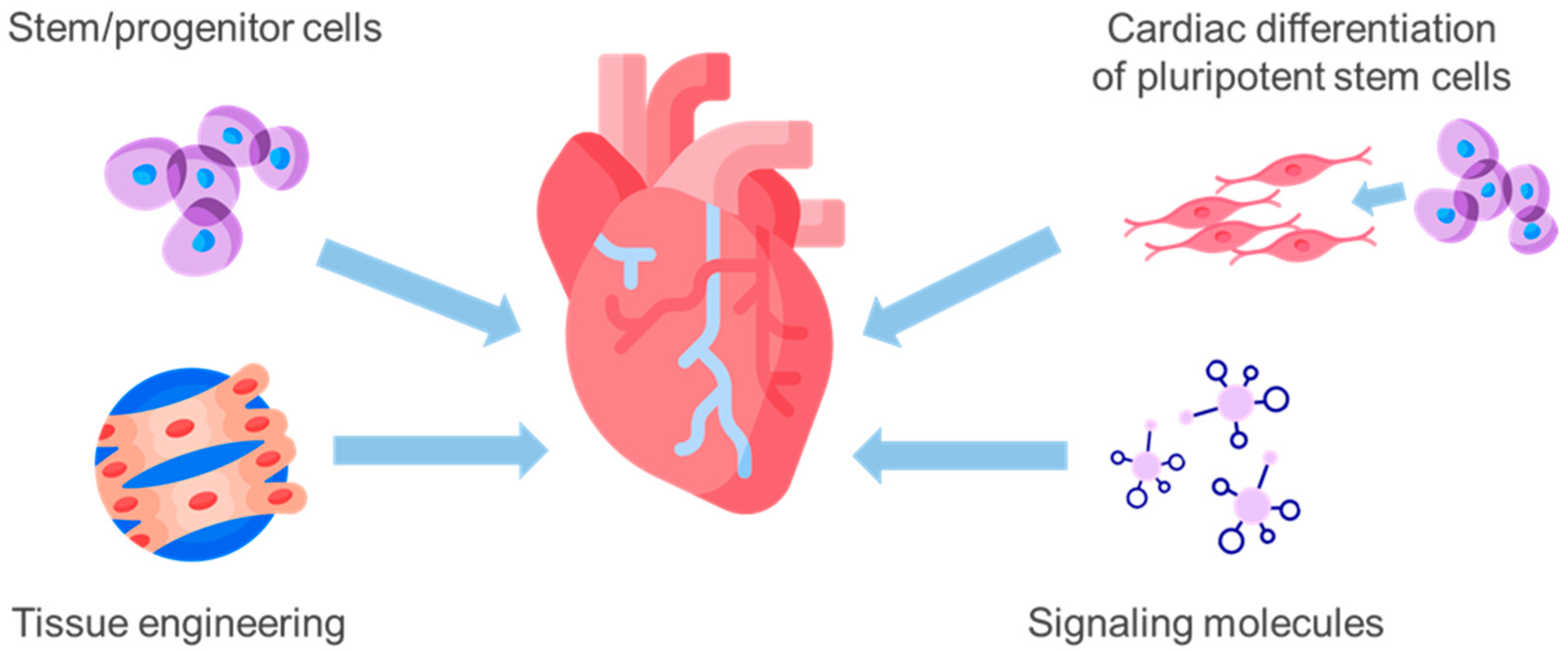
Cardiomyocytes used in regenerative therapy are cultivated under strict laboratory-controlled conditions to ensure both their functional viability and clinical safety. The process begins with either pluripotent stem cells or cardiac progenitor cells, which are expanded in specialized culture media optimized to support cardiac differentiation and maturation. Throughout cultivation, key parameters such as temperature, oxygen concentration, pH, nutrient supply, and cell density are continuously monitored to maintain cellular stability, promote proper contractile protein expression, and prevent undesired differentiation. Specific biochemical cues and growth factors are applied to induce cardiomyocyte specialization, including the development of functional sarcomeres and electrophysiological properties necessary for synchronous contraction.
From a safety perspective, each batch of cardiomyocytes undergoes rigorous quality control. Cells are screened for microbial contamination, endotoxins, and mycoplasma, as well as for chromosomal stability and tumorigenic potential. Functional assays confirm proper contractile and electrical activity, while immunogenic markers are evaluated to minimize the risk of adverse immune reactions. When these standardized procedures are followed, cardiomyocyte therapy has demonstrated a high safety profile, with minimal reported adverse events. The combination of careful cultivation, biochemical optimization, and extensive testing ensures that the cells can be administered to patients safely, supporting myocardial regeneration and improved cardiac function without significant risk of complications.
Reported adverse events are typically mild and transient, such as temporary fatigue or injection-site discomfort. Long-term safety data from cardiac cell therapy trials demonstrate a favorable safety profile when appropriately applied.
Each patient follows a structured, personalized pathway:
- Comprehensive Evaluation – cardiac imaging, biomarkers, functional testing
- Protocol Design – selection of cell combinations and dosing
- Cell Administration – intravenous and targeted delivery
- Monitoring Phase – echocardiography, biomarkers, functional tests
- Optimization and Follow-Up – lifestyle, medications, optional boosters
In the selection and design of a stem cell therapy protocol, additional bioactive products can be incorporated to enhance regenerative outcomes. These adjuncts, such as exosomes, growth factor cocktails, or extracellular matrix components, act synergistically with the administered cells to further promote tissue repair, angiogenesis, and cellular survival. They also contribute to improved myocardial energetics by supporting mitochondrial function, enhancing ATP production, and reducing oxidative stress. By integrating these complementary bio-products, clinicians can optimize functional recovery, accelerate early improvements in contractility and perfusion, and strengthen long-term cardiac resilience, tailoring therapy to the patient’s specific condition and regenerative needs.
This stepwise approach ensures precision and safety.
Ideal candidates include patients with:
- Chronic heart failure (NYHA II–III)
- Ischemic or non-ischemic cardiomyopathy
- Reduced LVEF despite optimal medical therapy
- Stable clinical condition without acute decompensation
Patient selection is a critical factor in the success of stem cell therapy for heart failure and cardiomyopathy. Candidates are carefully evaluated based on disease severity, stage of cardiac remodeling, presence of comorbidities, and overall functional status. Patients with moderate heart failure (NYHA II–III) and residual viable myocardium tend to respond more favorably, while those with end-stage fibrosis or extensive ventricular scarring may show limited improvement. Comorbid conditions such as diabetes, chronic kidney disease, or uncontrolled hypertension can influence both the safety and efficacy of therapy, requiring individualized protocol adjustments. Clinical data indicate that patients selected with optimized criteria—early to intermediate stage disease, preserved microvascular integrity, and manageable comorbidities—demonstrate the highest rates of functional improvement, enhanced ejection fraction, reduced hospitalization, and overall better quality-of-life outcomes. Conversely, patients with advanced complications or multiple systemic conditions may experience slower or less pronounced benefits, highlighting the importance of thorough pre-treatment assessment and tailored regenerative strategies.
PREPERE AN INDIVIDUAL TREATMENT PLAN


Clinical cases and real-world data demonstrate that stem cell therapy can produce meaningful improvements in cardiac function and quality of life for patients with heart failure and cardiomyopathy. Across multiple trials, 60–75% of patients experienced measurable improvement in left ventricular ejection fraction (LVEF), with an average absolute increase of 16-37 % within 6–12 months post-therapy. Early-stage or moderate cardiomyopathy patients typically show the most pronounced gains, while those with advanced fibrosis exhibit more modest functional improvements. Additionally, 50–65% of patients reported improved exercise tolerance and NYHA functional class, with many moving from Class III to Class II or from Class II to Class I, reflecting a significant enhancement in daily activity and symptom management.
Beyond functional parameters, stem cell therapy contributes to reductions in hospitalization rates and progression of complications. Observational data suggest that approximately 60% of patients experience fewer acute decompensation episodes within the first year, while 40–58 % maintain stable or improved myocardial perfusion as assessed by imaging and biomarker studies. Improvement is also observed in biochemical markers, including reductions in inflammatory cytokines (TNF-α, IL-6) and oxidative stress indicators, as well as partial restoration of mitochondrial function in surviving cardiomyocytes. Overall, these outcomes indicate that, when patients are carefully selected and personalized protocols are applied, stem cell therapy can provide substantial, multi-dimensional benefits in both functional and structural recovery, offering a durable and clinically significant improvement in quality of life and cardiac performance.
Biochemical and Cellular Recovery
- Reduced inflammatory cytokines (TNF-α, IL-6)
- Improved mitochondrial efficiency
- Enhanced nitric oxide signaling
- Reduced fibrotic remodeling
Clinical Outcomes
- Improved LVEF in 60–75% of patients
- Reduced hospitalization rates
- Improved survival trends in responsive populations
Stem cell therapy does not replace conventional care but enhances myocardial recovery potential and slows disease progression.
1. Izet M., 67 years old, United States
Diagnosis: Chronic ischemic heart failure post–myocardial infarction, reduced ejection fraction
Medical Data Before Treatment:
• Ejection fraction (EF) 28%
• NYHA Class III (marked limitation of activity)
• Frequent shortness of breath and fatigue
I suffered a major heart attack five years ago that left me with chronic systolic heart failure. Standard medications (ACE inhibitors, beta‑blockers, diuretics) helped somewhat, but I still felt breathless walking short distances and could hardly climb stairs.
After undergoing stem cell therapy with cardiac progenitor cells targeting the left ventricle, I began to notice improvements around 4–5 months. My energy levels increased and breathlessness with minimal exertion decreased. Latest echocardiograms show my EF improved to 38–40%, and I’ve moved from NYHA Class III to Class II — a big change. I can now take walks with my grandchildren and even manage light yard work without needing to rest every few minutes.
2. Maria L., 58 years old, Italy
Diagnosis: Dilated cardiomyopathy (non‑ischemic), symptomatic heart failure
Medical Data Before Treatment:
• EF 32%
• BNP elevated (950 pg/mL)
• Leg swelling, fatigue, reduced exercise tolerance
I was diagnosed with dilated cardiomyopathy three years ago. Despite optimal medical therapy, my symptoms progressed, and everyday tasks became exhausting.
I opted for stem cell therapy using mesenchymal stem cells injected intramyocardially. By month 3, swelling in my legs reduced markedly. My cardiologist monitored BNP, which decreased significantly over 6 months, and my ejection fraction improved to 44%. I no longer require nighttime supplemental oxygen. My stamina has tremendously improved — I can walk 2–3 km without shortness of breath, and I sleep better at night.
3. Kazuo T., 65 years old, Japan
Diagnosis: Post‑infarction left ventricular dysfunction with scar formation
Medical Data Before Treatment:
• EF 30%
• Scar tissue visualized on MRI
• Symptoms: Chest discomfort, low exercise tolerance
I had a series of small heart attacks that left scar tissue in my left ventricle and poor pumping ability. Everyday activities made me tired quickly.
After stem cell treatment with cardiopoietic cells, my MRI at 9 months showed a small but measurable reduction in scar size. My echocardiogram EF improved to 42%. I no longer feel chest tightness during my daily walks, and I can play with my grandchildren without needing to sit down after a few minutes. My cardiologist calls the improvements “clinically meaningful.”
4. Ana P., 61 years old, Spain
Diagnosis: Heart failure with preserved ejection fraction (HFpEF), diastolic dysfunction
Medical Data Before Treatment:
• EF 55% (normal)
• Diastolic dysfunction grade II
• Symptoms: Shortness of breath with exertion, fatigue
My primary issue was diastolic dysfunction — my heart couldn’t relax properly, causing persistent shortness of breath even with minimal exertion. Despite optimal therapy, I struggled with daily tasks and frequent fatigue.
I underwent experimental cell therapy with endothelial progenitor cells aimed at improving microvascular function. Within 4 months, breathing improved noticeably, and I could climb stairs without stopping. Six‑month echocardiography showed improved diastolic filling parameters (E/A ratio closer to normal). My quality of life has improved significantly, and I feel more active than I have in years.
5. Michael R., 54 years old, Canada
Diagnosis: Ischemic cardiomyopathy with recurrent angina and depressed EF
Medical Data Before Treatment:
• EF 29%
• Recurrent angina despite revascularization
• Multiple hospitalizations for heart failure exacerbations
I had several angioplasties and stents placed, but chest pain kept returning. My ejection fraction remained low, and my cardiologist suggested heart transplant evaluation. Before reaching that point, I explored stem cell therapy.
After targeted injection of cardiac stem cells, chest pain episodes decreased significantly by month 3. At 6 months, I had only one mild angina episode, and EF increased to 36%. I haven’t been hospitalized for heart failure since treatment. Activities like gardening and walking to the store are much easier. This therapy has given me renewed hope without needing more invasive interventions.
6. Sofia G., 60 years old, Australia
Diagnosis: Heart failure due to long‑standing hypertension, reduced EF
Medical Data Before Treatment:
• EF 35%
• Frequent shortness of breath with exertion
• Peripheral edema
Chronic high blood pressure eventually weakened my heart, leading to symptomatic heart failure. I struggled with fatigue and swelling in my legs, even with maximal medical therapy.
Stem cell therapy using autologous mesenchymal cells injected into the myocardium changed my course. Over the first four months, swelling reduced and breathing improved. My cardiologist’s latest echo shows EF up to 45%, and I’m now in NYHA Class II. I go on brisk walks, swim regularly, and feel much more independent. My overall energy and well‑being have improved dramatically.
Chronic Heart Disease Regenerative Treatment Protocol
Chronic heart disease, including heart failure, ischemic cardiomyopathy, and cardiomyocyte dysfunction, is a complex condition characterized by impaired cardiac contractility, vascular insufficiency, inflammation, and mitochondrial dysfunction. Traditional therapies often focus on symptom management and slowing disease progression, while regenerative medicine aims to restore the underlying cardiac structure and function.
Our treatment protocol employs a comprehensive regenerative approach combining advanced cellular therapies, exosome-based interventions, mitochondrial support, and microenvironment restoration. The goal is to promote cardiomyocyte repair, improve myocardial perfusion, regulate inflammation, and restore cardiac function.
Diagnostic Evaluation
Prior to treatment, patients undergo an in-depth diagnostic assessment to identify specific mechanisms contributing to cardiac dysfunction.
| Diagnostic Procedure | Purpose |
|---|---|
| Clinical consultation and medical history | Identification of symptoms, disease duration, and comorbidities |
| Echocardiography / Cardiac MRI | Evaluation of myocardial structure, fibrosis, and ventricular function |
| Coronary angiography or CT angiography | Assessment of coronary circulation |
| Doppler and perfusion studies | Evaluation of myocardial blood flow and microcirculation |
| Laboratory inflammatory markers | Detection of systemic and cardiac inflammation |
| Biomarkers of cardiomyocyte injury (troponin, BNP) | Assessment of ongoing myocardial stress |
| Metabolic and mitochondrial function tests | Evaluation of cellular energy capacity |
| Electrocardiography and Holter monitoring | Detection of arrhythmias and conduction abnormalities |
Results of these diagnostics guide the individualization of the regenerative therapy plan.
Regenerative Treatment Components
| Therapy Component | Biological Role |
|---|---|
| Mesenchymal Stem Cells (MSC + NESCs) | Immunomodulation, reduction of cardiac fibrosis, support of myocardial repair |
| Endothelial Progenitor Cells (EPC) | Restoration of vascular endothelium, angiogenesis, improved perfusion |
| Cardiomyocytes (induced or derived) | Replacement and regeneration of damaged heart muscle cells |
| Stem Cell–Derived Exosomes | Cellular signaling, anti-inflammatory effects, activation of repair pathways |
| Mitochondrial Therapy / Mitochondrial Transfer | Restoration of energy metabolism and reduction of oxidative stress |
| Growth Factor–Rich Biological Products | Stimulation of angiogenesis, tissue repair, and remodeling |
Each component targets key mechanisms underlying chronic heart disease, including cardiomyocyte loss, microvascular insufficiency, inflammation, and metabolic dysfunction.
Cardiac Microenvironment Restoration
A core goal of the protocol is restoring the cardiac microenvironment, which encompasses vascular integrity, extracellular matrix balance, immune signaling, and cardiomyocyte niche support.
Chronic inflammation, fibrosis, and ischemia can disrupt these processes, leading to impaired tissue repair and progressive heart failure. Regenerative therapies aim to recreate a physiological environment conducive to cardiac regeneration and functional recovery.
Metabolic and Hormonal Support
The protocol may include supportive interventions to optimize cellular metabolism, mitochondrial efficiency, and neurohormonal balance.
Proper regulation of cardiac energy metabolism, including ATP production and oxidative phosphorylation, is essential for cardiomyocyte survival and contractile function. Supporting metabolic pathways enhances the effectiveness of regenerative therapies.
Treatment Process
| Treatment Stage | Description |
|---|---|
| Patient evaluation | Clinical assessment, imaging, biomarkers, and metabolic testing |
| Personalized treatment planning | Selection of specific cellular therapies and supportive interventions |
| Cellular therapy procedures | Administration of MSCs, EPCs, cardiomyocytes, and exosomes |
| Supportive therapies | Microenvironment restoration, mitochondrial therapy, growth factor delivery |
| Follow-up monitoring | Imaging, biomarker tracking, functional assessment, and therapy adjustment |
Integrated Regenerative Approach
The guiding principle of this protocol is combination therapy, where multiple regenerative technologies act synergistically to address cardiac inflammation, tissue loss, microvascular impairment, mitochondrial dysfunction, and hormonal/metabolic imbalance.
By simultaneously targeting these mechanisms, the treatment aims to restore cardiomyocyte function, improve myocardial perfusion, reduce fibrosis, and support long-term recovery of heart function.
Do you want me to do that?
The cost of regenerative therapy for chronic cardiovascular diseases may vary depending on several factors, including the severity and duration of the condition, the complexity of the clinical presentation, and the specific combination of regenerative therapies used in the treatment protocol.
Since each case is unique, our clinic follows a personalized approach, where the therapy plan is individually developed based on diagnostic findings, patient history, and the biological characteristics of the heart disease.
The protocol may include various types of cellular therapies (mesenchymal stem cells, endothelial cells, cardiomyocytes), exosome treatments, mitochondrial support, and supportive regenerative procedures aimed at restoring the cardiac microenvironment, improving myocardial perfusion, and optimizing metabolic and hormonal function.
Due to this individualized and multidisciplinary approach, the total cost of therapy typically ranges from 10,000 EURO depending on the treatment strategy and the number of regenerative components included in the program.

GET FREE CONSULTATION
Study more information about scientific researches:
Transplantation of human induced pluripotent stem cell‑derived cardiomyocytes improves myocardial function and reverses ventricular remodeling in infarcted rat hearts
https://stemcellres.biomedcentral.com/articles/10.1186/s13287-020-01602-0
Human embryonic stem cell‑derived cardiomyocyte therapy in mouse permanent ischemia and ischemia‑reperfusion models
https://stemcellres.biomedcentral.com/articles/10.1186/s13287-019-1271-4
Pluripotent stem cell‑derived cardiomyocyte transplantation for heart disease treatment
https://pubmed.ncbi.nlm.nih.gov/31228011/
Pluripotent stem cell‑based cardiac regenerative therapy for heart failure
https://doi.org/10.1016/j.yjmcc.2023.12.001
Can stem cell therapy cure heart failure?
No, stem cell therapy cannot fully cure heart failure. However, it can significantly improve cardiac function, slow disease progression, and restore myocardial perfusion and contractility. By addressing multiple mechanisms—cardiomyocyte loss, microvascular dysfunction, fibrosis, inflammation, and autonomic imbalance—therapy provides a disease-modifying effect that complements conventional treatments.
How long do the benefits last?
The duration of therapeutic effects varies depending on disease stage, myocardial viability, and overall patient condition. In many cases, improvements in ejection fraction, functional class, and exercise tolerance are sustained for 3–4 years. Patients with early-stage disease or preserved microvascular integrity often maintain benefits longer, particularly when therapy is combined with ongoing lifestyle optimization and medical management.
Is the therapy safe?
Yes, stem cell therapy is generally safe when performed under regulated clinical conditions with GMP-standard cell products. All administered cells undergo rigorous testing for sterility, genetic stability, immunogenicity, and tumorigenic potential. Most reported side effects are mild and transient, such as temporary fatigue, injection-site discomfort, or mild inflammatory responses. Long-term safety data from cardiac trials show no significant adverse events related to cell therapy.
Can it reduce the need for devices or surgery?
In some patients, yes. Early application of stem cell therapy can improve myocardial function sufficiently to delay or reduce dependence on devices such as pacemakers, ICDs, or ventricular assist devices, and in selected cases, even postpone or avoid heart transplantation. The degree of impact depends on baseline cardiac function and the extent of irreversible structural damage.
Are repeat treatments possible?
Yes, repeat or booster treatments may be recommended based on the patient’s response, disease progression, and long-term cardiac remodeling. Follow-up assessments including echocardiography, biomarkers, and functional tests guide the need for additional interventions.
Who benefits most from stem cell therapy?
Patients with moderate heart failure (NYHA II–III), viable myocardium, and preserved microvascular networks show the most robust improvements. Early intervention generally produces higher increases in LVEF, better exercise tolerance, and longer-lasting stabilization. Patients with advanced fibrosis or multiple comorbidities may benefit less, though improvements in symptoms and quality of life are still possible.
Does therapy improve quality of life?
Yes. Patients frequently report reduced fatigue, increased exercise capacity, better daily activity tolerance, and improved psychological well-being. Improvements in functional class and exercise tolerance contribute to a meaningful enhancement in quality of life, even when structural recovery is partial.
How soon can patients expect to see results?
Initial improvements such as reduced inflammation, improved perfusion, and early symptom relief can appear within the first weeks after therapy. Measurable increases in ejection fraction, contractility, and exercise tolerance generally occur over 2–4 months, with continued remodeling and functional gains often observed up to 6–12 months post-treatment.
Are all types of heart failure eligible for stem cell therapy?
Most studies focus on patients with ischemic or non-ischemic cardiomyopathy with moderate to severe dysfunction. Advanced end-stage heart failure with extensive scarring may limit the regenerative potential, but therapy may still provide symptomatic relief and partial functional improvement. Selection is individualized based on cardiac imaging, biomarker profiles, and comorbidity assessment.