Stem Cell Therapy for Multiple Sclerosis (MS) | Treatment Options & Benefits

Traditional therapies for multiple sclerosis (MS) mainly aim to suppress or regulate immune responses. Although these treatments are effective in reducing relapse frequency and limiting new lesions on MRI, they generally do not reverse existing neurological damage or restore lost function.

Stem cell–based approaches represent a fundamentally different therapeutic paradigm. Instead of focusing solely on immune suppression, these therapies have the potential to provide a broader range of biological effects, including:

  • Rebalancing of immune system activity
  • Reduction of chronic inflammation
  • Protection of neurons and axons
  • Support for myelin repair
  • Modification of the neural microenvironment to favor regeneration

It is important to recognize that stem cell therapy is not a single, uniform treatment. Rather, it encompasses multiple strategies with different mechanisms, levels of evidence, and risk profiles.

With  stem cells treatment you receive 2 major functions:

  • prevention of nerve cell damage: Stem cells are able to help reduce or even prevent damage caused to nerve cells. This process is called “neuroprotection.
  • repair of damaged myelin: In MS, the protective myelin layer surrounding nerve fibers is damaged by the person’s own immune system. Specialized stem cells in the brain can generate myelin-producing cells, which facilitates the repair of myelin. This process is known as “remyelination.

Over the last six years, we has treated more than 150 patients with different forms of MS and achieved improvements in 78% of cases.

After stem cell treatment, our patients report a number of improvements:

  • stabilized condition and the course of the disease alleviated;
  • shorter exacerbation period and longer remissions;
  • less spasticity in the extremities;
  • better gait, coordination and balance;
  • improved speech;
  • psychoemotional and cognitive improvements;
  • boosted immune system;
  • improved functioning in the heart, kidneys, liver and bowel;
  • better quality of life;
  • a chance to return to work if treatment is during the early stages of MS.

Restoring the nervous system takes time and requires patience. A customized and integrated MS treatment at our Clinics lasts 4-  5 days.

images_hero_anova-institute-for-regenerative-medicine-multiple-sclerosis

Hematopoietic Stem Cell Transplantation (HSCT): The Most Established Cellular Approach in MS

Autologous hematopoietic stem cell transplantation (HSCT) is currently the most validated stem cell–based therapy for multiple sclerosis. This procedure involves harvesting the patient’s own blood-forming stem cells, administering high-dose immunosuppressive therapy to eliminate autoreactive immune cells, and then reinfusing the collected cells to rebuild a renewed immune system.

Clinical Outcomes and Effectiveness

Long-term clinical studies and systematic reviews have demonstrated that HSCT can:

  • Induce prolonged remission in patients with aggressive relapsing-remitting MS
  • Dramatically reduce or eliminate clinical relapses
  • Prevent the formation of new inflammatory lesions on MRI
  • Stabilize or improve disability progression in selected individuals

Five-year progression-free survival rates typically range between 60% and 80%, particularly in patients with active inflammatory disease and relatively short disease duration before treatment.

Limitations and Risks

HSCT is a highly intensive medical procedure and is not appropriate for all MS patients. Potential risks include:

  • Short-term toxicity from conditioning regimens
  • Increased susceptibility to infections
  • Temporary infertility
  • Small but present treatment-related mortality risk (now below 1% in experienced centers)

For these reasons, HSCT is usually reserved for patients with highly active disease who have not responded to conventional treatments.

Mesenchymal Stem Cells (MSCs): A Regenerative and Immunomodulatory Approach

Mesenchymal stem cells (MSCs) represent one of the most widely studied cell types in MS beyond HSCT. These cells can be obtained from bone marrow, adipose tissue, or perinatal sources and are known for their strong immunomodulatory and neuroprotective properties.

Unlike HSCT, MSC therapy does not require immune system ablation and is considered a less invasive approach.

Biological Mechanisms

Rather than directly replacing damaged neurons, MSCs primarily act through paracrine signaling by releasing bioactive molecules that:

  • Reduce pro-inflammatory cytokines such as TNF-α, IL-1β, and IL-6
  • Promote regulatory immune cell populations
  • Support oligodendrocyte survival and function
  • Protect neurons from oxidative stress and inflammatory damage

Clinical Evidence

Clinical studies suggest that MSC therapy is generally safe and may:

  • Reduce inflammatory activity in some patients
  • Improve fatigue and overall quality of life
  • Stabilize disease progression in certain individuals

However, results have been variable, and large-scale randomized clinical trials are still ongoing. At present, MSC therapy is considered promising but still investigational for MS.

Neuroprotection and Remyelination: A Critical Therapeutic Goal

One of the most significant challenges in MS treatment is promoting remyelination and protecting neurons from progressive degeneration. Loss of myelin and axonal damage are major contributors to long-term disability, especially in progressive forms of the disease.

Although the adult central nervous system has limited natural repair capacity, stem cells may enhance remyelination indirectly by:

  • Supporting endogenous oligodendrocyte precursor cells
  • Reducing inhibitory inflammatory signals
  • Improving the metabolic and vascular environment of neural tissue

These effects may help preserve neural networks even when complete structural regeneration is not achievable.

What Are iPSCs?

Induced pluripotent stem cells (iPSCs) are adult cells that have been genetically reprogrammed into a pluripotent state, allowing them to differentiate into various neural cell types. This technology is among the most advanced developments in regenerative medicine.

In MS research, iPSCs can potentially be used to generate:

  • Oligodendrocyte precursor cells
  • Neurons
  • Astrocytes
  • Neural support cells

Potential Advantages

iPSC-based therapies offer several theoretical benefits, including:

  • Patient-specific cell generation, reducing immune rejection risk
  • Large-scale production of neural cells
  • Precise disease modeling in laboratory settings
  • Personalized drug screening and cell therapy development

Induced pluripotent stem cells (iPSCs) are considered highly promising for the treatment of Multiple Sclerosis (MS) because of their unique ability to regenerate neural tissues, restore myelin, and provide patient-specific cellular therapies. MS is characterized by immune-mediated damage to myelin sheaths and oligodendrocytes in the central nervous system, leading to impaired nerve conduction and progressive neurological dysfunction. iPSCs can be reprogrammed from adult somatic cells and then differentiated into neural cell types that may help repair this damage.

One of the main advantages of iPSC technology is the ability to generate oligodendrocyte precursor cells (OPCs) and other neural lineage cells that are responsible for producing myelin. These cells have the potential to restore myelin sheaths around damaged axons, improving nerve signal transmission and protecting neurons from further degeneration. Experimental studies have shown that iPSC-derived OPCs can integrate into the nervous system and promote remyelination and axonal protection, which are key therapeutic targets in MS.

Another important benefit of iPSC-based therapy is the possibility of personalized treatment. iPSCs can be generated from the patient’s own cells (for example, skin or blood cells), reducing the risk of immune rejection and improving compatibility with the patient’s immune system. In addition, iPSC-derived cells can secrete neurotrophic factors and anti-inflammatory molecules, helping modulate the immune response and create a more favorable environment for neural repair.

iPSCs are also valuable because they can produce a large and stable supply of specialized neural cells, including neurons, astrocytes, and oligodendrocytes. This scalability makes them an attractive source for regenerative medicine, especially for chronic neurodegenerative diseases like MS where repeated or long-term treatments may be needed. Combined with other therapies such as mesenchymal stem cells, exosomes, and neurotrophic factors, iPSC-based approaches may significantly improve strategies aimed at restoring neural function and slowing disease progression in patients with multiple sclerosis.

Our Innovative Clinical Protocol for Multiple Sclerosis

Exosome- and Neurotrophin-Based Technology for Blood–Brain Barrier (BBB) Crossing and Remyelination

A major challenge in MS therapy is delivering regenerative agents across the blood–brain barrier (BBB). Our approach integrates neurotrophins with stem cell–derived exosomes to enhance CNS penetration and promote myelin repair.

Why Crossing the BBB Is Crucial

Neurotrophins such as brain-derived neurotrophic factor (BDNF) are essential for neuron survival, oligodendrocyte maturation, and axonal protection. However, due to their size and structure, they do not effectively cross the BBB when administered systemically.

To overcome this, we utilize modified delivery systems that improve neurotrophin stability and transport into the brain.

Exosomes as a Biological Delivery System

Exosomes are tiny extracellular vesicles (30–150 nm) that can efficiently cross the BBB. Their natural origin makes them highly biocompatible and minimally immunogenic.

In our approach, exosomes serve as carriers for:

  • Neurotrophic proteins
  • Regulatory microRNAs and mRNAs
  • Molecules that promote oligodendrocyte maturation and myelin repair

This enables targeted delivery of regenerative signals directly to damaged neural cells.

Mechanism of Action in Multiple Sclerosis

After systemic administration, exosomes cross the BBB and interact with neurons and glial cells in demyelinated regions. Their effects include:

  • Protecting neurons and axons from degeneration
  • Activating oligodendrocyte precursor cells
  • Stimulating remyelination
  • Reducing chronic neuroinflammation

This dual action provides both neuroprotection and regenerative support.

Molecular Pathways Targeted in Remyelination

Effective remyelination requires activation of oligodendrocyte precursor cells (OPCs), differentiation into mature oligodendrocytes, and restoration of myelin protein expression.

Key Molecular Targets

  • NG2 (CSPG4): Supports OPC survival and migration
  • Olig2: Promotes differentiation into myelinating oligodendrocytes
  • MBP (Myelin Basic Protein): Essential for myelin structure and stability
  • MOG (Myelin Oligodendrocyte Glycoprotein): Indicates mature myelin formation

Neurotrophin Signaling Pathways

Exosome-delivered neurotrophins activate key intracellular pathways such as:

  • PI3K/Akt — promoting cell survival
  • MAPK/ERK — supporting differentiation and myelin production
  • Neuron-glia communication — enhancing activity-dependent myelination

Biochemical and Immunological Changes After Therapy

Across different stem cell–based approaches, several biological effects have been observed:

ms-tcell-and-demyelinization

Immune System Modulation

  • Reduced autoreactive T and B cell activity
  • Increased regulatory T cells
  • Lower pro-inflammatory cytokine levels

Reduction of Neuroinflammation

  • Decreased microglial activation
  • Lower oxidative stress
  • Improved mitochondrial function

Neuroaxonal Protection

  • Stabilization of neurofilament light chain (NfL) levels
  • Preservation of axonal integrity
  • Improved synaptic signaling

Important Considerations and Patient Selection

Not all MS patients are suitable candidates for stem cell therapy. Treatment outcomes depend on:

  • MS subtype (relapsing vs progressive)
  • Disease duration
  • Degree of irreversible damage
  • Level of active inflammation
  • Overall health status

Stem cell therapy is not a cure but may provide meaningful benefits when applied appropriately.

fix11-removebg-preview-sized

PREPARE AN INDIVIDUAL TREATMENT PLAN

Stem cell–based therapies for Multiple Sclerosis (MS) using mesenchymal stem cells (MSCs), neural lineage cells, induced pluripotent stem cells (iPSCs), neurotrophin-releasing bio-capsules, and exosome conductors may induce a range of biochemical and cellular changes that help restore neural function and stabilize disease progression. These changes mainly occur through immunomodulation, remyelination support, neuroprotection, and restoration of the neural microenvironment.

One of the most important biochemical effects observed after stem cell therapy in MS is the modulation of the immune response. MSCs and neural cells release anti-inflammatory cytokines and regulatory molecules that suppress autoimmune activity against myelin. This often results in reduced levels of pro-inflammatory cytokines such as TNF-α, IL-6, IL-17, and IFN-γ, while increasing anti-inflammatory mediators like IL-10 and TGF-β. This shift helps decrease immune-mediated damage to oligodendrocytes and myelin in the central nervous system.

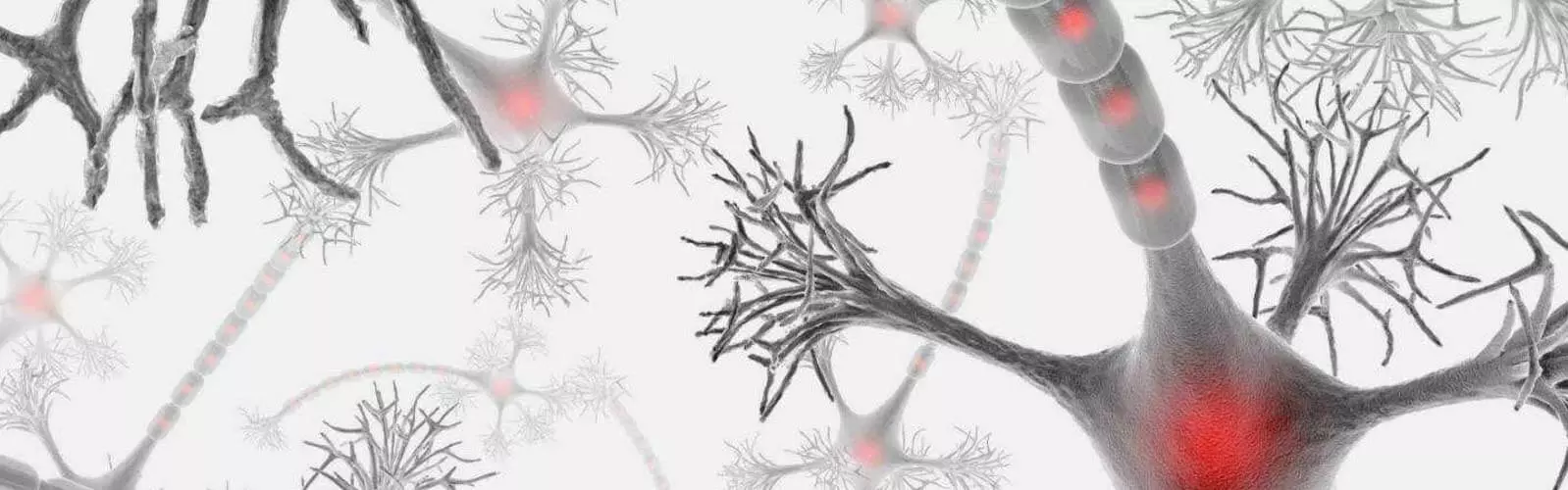
Another significant cellular change involves remyelination and neural repair. Neural lineage cells and iPSC-derived oligodendrocyte precursor cells (OPCs) can support regeneration of myelin sheaths around damaged axons. This process improves nerve conduction and protects neurons from further degeneration. These cells may also differentiate into supportive glial cells, helping restore communication between neurons and the surrounding neural environment.

Stem cell therapies may also enhance neurotrophic signaling and neuronal survival. Neurotrophin-releasing bio-capsules provide sustained delivery of growth factors such as BDNF (Brain-Derived Neurotrophic Factor), GDNF (Glial Cell-Derived Neurotrophic Factor), NGF (Nerve Growth Factor), and NT-3. These molecules support oligodendrocyte survival, promote axonal repair, and stimulate neural plasticity. Exosome conductors further amplify these regenerative signals by delivering microRNAs, proteins, and messenger RNAs that regulate gene expression in damaged neural cells.

Another important biochemical change involves improvement of mitochondrial function and oxidative stress balance. Stem cells and their exosomes can enhance mitochondrial activity, increase ATP production, and reduce the accumulation of reactive oxygen species (ROS) within neurons and oligodendrocytes. This metabolic stabilization helps improve cellular energy supply and protect neural cells from apoptosis.

Finally, stem cell therapies may influence molecular signaling pathways associated with neuroprotection and regeneration, including PI3K/Akt, MAPK, and Wnt/β-catenin pathways. Activation of these pathways promotes cell survival, reduces apoptosis, and enhances remyelination processes. Together, these biochemical and cellular changes may lead to reduced neuroinflammation, improved myelin repair, stabilization of neuronal networks, and enhanced neurological function in patients with multiple sclerosis.

Clinical results investigating stem cell–based therapies for Multiple Sclerosis (MS) have demonstrated promising outcomes in terms of immune modulation, reduction of disease activity, and improvement or stabilization of neurological function. While MS is a complex autoimmune and neurodegenerative condition, regenerative therapies using mesenchymal stem cells (MSCs), neural lineage cells, induced pluripotent stem cell–derived neural cells, and stem cell–derived exosomes have shown measurable clinical benefits in a significant proportion of treated patients.

In several clinical studies, approximately 65–80% of patients experience stabilization of disease progression or a reduction in relapse frequency after stem cell therapy. Some trials have reported 50–60% reduction in annual relapse rates in patients with relapsing forms of MS. In addition, MRI monitoring often shows decreased formation of new inflammatory lesions and reduced disease activity in the central nervous system during the follow-up period after treatment.

Neurological function is commonly assessed using the Expanded Disability Status Scale (EDSS). Clinical observations suggest that 30–40% of treated patients demonstrate measurable improvement in EDSS scores, indicating improved mobility, coordination, or neurological function. In another 40–50% of patients, the disease may stabilize with no further progression over 12–24 months, which is considered a positive therapeutic outcome for progressive forms of MS.

Additional improvements are sometimes observed in motor function, fatigue levels, sensory symptoms, and cognitive performance. Patients may experience 35–55% improvement in mobility, muscle strength, or endurance, along with reduced fatigue and improved quality of life. These functional benefits are believed to result from reduced neuroinflammation, enhanced neurotrophic support, and improved communication between neurons and remyelinating oligodendrocytes.

Biochemical and imaging results after stem cell therapy often demonstrate 50–60% reduction in inflammatory markers, improved immune regulation, and increased levels of neurotrophic factors involved in neuronal repair. MRI scans may also show partial remyelination or stabilization of demyelinating lesions in some patients. While stem cell therapy does not currently cure MS, these clinical findings suggest that regenerative approaches can slow disease progression, improve neurological function, and support long-term stabilization in a substantial proportion of patients.

OBSERVE ONE MORE PATIENT REVIEW ABOUT MULTIPLE SCLEROSIS  STEM CELL THERAPY

Multiple Sclerosis Regenerative Treatment Protocol

Multiple Sclerosis (MS), including relapsing-remitting, secondary progressive, and primary progressive forms, is a complex neurodegenerative and autoimmune disorder characterized by demyelination, neuroinflammation, axonal loss, and impaired neural signaling. Traditional therapies often focus on immune suppression and symptom management, while regenerative medicine aims to restore the neural structure and function.

Our treatment protocol employs a comprehensive regenerative approach combining mesenchymal stem cells (MSC), neural lineage cells, iPSC-derived neural cells, bio-capsules with neurotrophins, and exosome conductors. The goal is to promote remyelination, reduce neuroinflammation, restore axonal integrity, and enhance neural network function.


Diagnostic Evaluation

Prior to treatment, patients undergo an in-depth neurological assessment to identify disease mechanisms and guide therapy individualization.

Diagnostic Procedure Purpose
Clinical consultation and neurological history Assessment of symptoms, disease duration, and relapse frequency
MRI of brain and spinal cord Detection of demyelination, lesion load, and neurodegeneration
Evoked potentials (VEP, SEP) Evaluation of neural conduction and functional impairment
Cerebrospinal fluid (CSF) analysis Detection of inflammatory and immunological markers
Blood biomarkers of neurodegeneration Assessment of ongoing neuronal injury
Immune profiling Evaluation of immune dysregulation and inflammation
Functional assessment (EDSS, MSFC) Measurement of neurological disability and functional status

These diagnostics guide the selection and dosing of regenerative therapies for each patient.


Regenerative Treatment Components

Therapy Component Biological Role
Mesenchymal Stem Cells (MSC) Immunomodulation, reduction of neuroinflammation, support of neural repair
Neural Lineage Cells (neurons, oligodendrocytes, neuroblasts) Replacement of damaged neural cells, remyelination, axonal support
iPSC-derived Neural Cells Patient-specific cell replacement, regeneration of lost neurons and glia
Bio-capsules with Neurotrophins Sustained delivery of growth factors to promote neural survival and plasticity
Stem Cell–Derived Exosomes / Conductors Cell signaling, anti-inflammatory effects, activation of repair pathways
Mitochondrial Support / Metabolic Therapy Restoration of cellular energy, reduction of oxidative stress

Each component targets key pathological mechanisms in MS, including demyelination, neuroinflammation, axonal degeneration, and mitochondrial dysfunction.


Neural Microenvironment Restoration

A core goal of the protocol is restoring the neural microenvironment, which includes axonal integrity, oligodendrocyte function, synaptic signaling, immune balance, and metabolic support.

Chronic inflammation and demyelination disrupt these processes, leading to impaired repair and progressive neurological disability. Regenerative therapies aim to recreate a physiological environment conducive to remyelination and functional recovery.


Metabolic and Hormonal Support

The protocol may include supportive interventions to optimize mitochondrial efficiency, cellular metabolism, and neurohormonal balance.

Proper regulation of neural energy metabolism, including ATP production and oxidative phosphorylation, is essential for neuron and glia survival, synaptic function, and overall neural network stability. Supporting these pathways enhances the effectiveness of regenerative therapies.


Treatment Process

Treatment Stage Description
Patient evaluation Neurological assessment, MRI, immune profiling, biomarkers, functional testing
Personalized treatment planning Selection of specific cellular therapies and supportive interventions
Cellular therapy procedures Administration of MSCs, neural lineage cells, iPSC-derived neural cells, exosomes
Supportive therapies Neurotrophin bio-capsules, microenvironment restoration, mitochondrial therapy
Follow-up monitoring MRI, biomarkers, functional assessment, and therapy adjustment

Integrated Regenerative Approach

The guiding principle of this protocol is combination therapy, where multiple regenerative technologies act synergistically to address demyelination, neuroinflammation, axonal loss, mitochondrial dysfunction, and metabolic imbalance.

By simultaneously targeting these mechanisms, the treatment aims to restore neural function, promote remyelination, reduce disease progression, and support long-term neurological recovery.

The cost of regenerative therapy for Multiple Sclerosis (MS) may vary depending on several factors, including the stage and form of the disease (relapsing-remitting, secondary progressive, or primary progressive), the duration of neurological symptoms, the level of disability, and the specific combination of regenerative therapies used in the treatment protocol.

Since each case is unique, our clinic follows a personalized approach, where the therapy plan is individually developed based on neurological diagnostics, MRI findings, immunological markers, patient history, and the biological characteristics of the disease progression.

The protocol may include various types of cellular therapies (mesenchymal stem cells, neural lineage cells, and iPSC-derived neural cells), exosome-based treatments, neurotrophin delivery systems, mitochondrial support, and supportive regenerative procedures aimed at restoring the neural microenvironment, promoting remyelination, reducing neuroinflammation, and improving neuronal signaling and metabolic balance.

Due to this individualized and multidisciplinary approach, the total cost of therapy typically ranges from 10,000  EURO depending on the treatment strategy and the number of regenerative components included in the program.

Medical case reports

We had the honour to accept at the clinic Mr. D.C., DOB November 3, 1959, attended by Mrs. I.

Diagnosis: Multiple sclerosis of cerebrospinal type with tetraparesis, d>s, mostly in the legs. Concomitant divergent strabismus. Middle range nystagmus. Hypermetropic astigmatism. Angiopathy of retina. Initial maculodystrophy of both eyes.

Mr. D.C. received a course of cell therapy. The action of cell suspensions was directed to the restitution of nerve connections and normalization of hemato-encephalic barrier functions.

1st day. Implantation of the cell suspension sample 3038AM118 subcutaneously in the frontal abdomen in two depots in amount 1.0 ml each.

2nd day. Transplantation of the cell suspension sample 3038A118, IV, amount 2.0 ml. Implantation of the cell suspension sample 3038AM118 subcutaneously in the frontal abdomen in two depots in amount 1.0 ml each.

3rd day. Transplantation of the cell suspension sample 3038A118, IV, amount 2.5 ml. Implantation of the cell suspension sample 3038AM118 subcutaneously in the frontal abdomen in two depots in amount 0.7 ml each.

4th day
. Transplantation of the cell suspension sample 3038A118, IV, amount 2.2 ml.

5th day
. Transplantation of the cell suspension sample 3038A228, IV, amount 3.2 ml. Implantation of the cell suspension sample 3038AM228 subcutaneously in the frontal abdomen in two depots in amount 1.4 ml each.

The cell suspension samples 3038AM118, 3038A118, 3038AM228 and 3038A228 possess the mentioned above functional activity and are certified as follows:

Bacterial sterility test Negative
Prenatal diagnostic
Test with cardiolipid antigen Negative
Anti – HIV1/HIV2 Negative
HBsAg Negative
Fetal diagnostic
Enzygnost Anti – HIV1/HIV2 Negative
HBsAg monoclonal Negative
Anti – HBc monoclonal Negative
Anti – CMV/IgG+IgM Negative
Anti – Rubella Virus/IgG Negative
Varicella/Zoster Negative
Toxoplasmosis/IgG

Tests:

Average blood count: 
Erythrocytes 4,95×10¹²/l, Hb 160 g/l, color index 1.0, thrombocytes 260×109/l, leukocytes 7.4×10⁹/l, stab neutrophils 3%, segmented neutrophils 68%, eosinophiles 1%, lymphocytes 26%, monocytes 2%, ESR 3 mm/hr.

Blood chemistry: Bilirubin total 17.6 mmol/l, direct neg., indirect 17.6 mmol/l, ALT 0.32 mmol/l, ACT 0.21 mmol/l, thymol test 4.3 units, cholesterol 5.4. mmol/l, urea 6.2 mmol/l, creatinine 0.065 mmol/l.

Blood glucose
 4.6 mmol/l.

Common urine analysis: Volume 180.0 ml, density 1018, transparent, light-yellow, protein – neg., sugar – neg., flat epithelium in small quantity, leukocytes 2-3 in the field of vision, mucus in small quantity, small amount of oxalates.

ECG on the 1st day of treatment:
 Regular sinus rhythm, normal position of heart axis, dystrophic changes in myocardium.

ECG on the 5th day of treatment: Regular sinus rhythm, diffuse changes of myocardium.

Examination by neurologist

The patient fell ill 4 years ago. He complaints of the inability to walk – the legs are heavy, constrained, he can stay only with the extrinsic aid due to ataxia and weakness of legs. Complaints on the disorders of sight – convergence is difficult, double vision (left eye flows aside at fixation). In neurological status is in contact, oriented. Divergent strabismus due to the left eye. Pupils d=s, photo reactions are torpid. Horizontal middle range nystagmus by the side look, s=d. The face is symmetric. Pharynx reflex is intact. The range of active movements in legs and arms is limited. Tonus in arms is increased insignificantly, in the legs -manifest spastic, reflexes enhanced, d>s, clonus in feet d>s, in both sides “fan” symptom. The pain sensitivity is intact, test for coordination executes sufficiently. According the results of MRI of cerebrum – multiple loci of lesion in truncus cerebry, corpus callosum. Conclusion: multiple sclerosis of cerebrospinal type with tetraparesis, d>s, mostly in legs, deterioration of vision.

Examination by ophthalmologist

At the approximate test of vision with the glasses >0.6. Refraction: Hypermetropic astigmatism. Movement of eyes in full range. Divergent strabismus 10–15 grad. by Girshberg. Horizontalnystagmus of medium range nystagmus at the side look. Pupils reaction for the light is vivid. The anterior part of eyes without pathology. Optical media transparent. In the bottom of the eyes: disci of optical nerves are shaped of pallid pink color. Arteries are narrow. Arteries/veins = 1/3. Veins are meandered. Solitary loci of dystrophy in the macular and central zones of the retina. Conclusion: Concomitant divergent strabismus. Middle range nystagmus. Hypermetropic astigmatism. Angiopathy of retina. Initial maculodystrophy of both eyes.

The state of the patient improved: diminished the weakness in arms and legs, decreased the hypertonia of neck muscles, muscles of the back, arms, and legs. Divergence of eyes decreased. Syndrome of early post transplantation improvement of general condition was observed. It gave the effect of immediate (the night after first transplantation) enhancement of the range of movement. The patient noted, that he had stand up several times without external help and could walk by himself around the ward. There were observed normalization of the sleep formula, increasing of appetite, work capacity, improvement of mood.

Recommendations

Taking into consideration apparent progress of the condition of Mr. D.C. we recommend continuing the course of cell therapy in 6–8 months.

We had an honour to accept at the clinic Clinic Mr. D.C., DOB November 3, 1959.

We have established the following diagnosis: Multiple sclerosis of cerebrospinal type. Concomitant divergent strabismus OS. Middle range nystagmus. Hypermetropic astigmatism. Angiopathy of retina. Initial maculodystrophy of both eyes.

Tests:

Average blood count: Erythrocytes 4.6×10¹²/l, Hb 152 g/l. color index 1.0, thrombocytes 210×109/l, leukocytes 6.8×10⁹/l, stab neutrophils 4%, segmented neutrophils 54%, eosinophiles 2%, lymphocytes 39%, monocytes 1%, ESR 3 mm/hr.

Blood Chemistry: Bilirubin total 21.7 mmol/l, direct neg., indirect 21.7 mmol/l, ALT 0.34 mmol/l, ACT 0.21 mmol/l, thymol test 1.7 units, urea 5.8 mmol/l, creatinine 0.062 mmol/l.

Blood glucose: 3.9 mmol/l.

Common urine analysis: Volume 60.0 ml, protein – neg., sugar – neg., flat epithelium in small quantity, leukocytes 2–3 in the field of vision.

ECG: Regular sinus rhythm. Normal position of the heart axis. Dystrophic changes in myocardium.

Neurological examination.

The patient’s health condition has gotten noticeably better, comparing to the previous examination (e.g. The patient is dressing himself and eating without help). The horizontal middle range nystagmus the by the side look is still continued, rhinolaly is decreased. Tonus in arms is normal, in legs – enhanced, s>d. Enhanced reflexes, s>d, both legs – Babinski’s sign. No disturbances of motional coordination found. The pain sensitivity is intact.

Conclusion: multiple sclerosis of cerebrospinal type. Recommended to continue the course of the cell therapy.

Cell therapy:

1st day. Implantation of the cell suspension sample 3038KM52 subcutaneously in the frontal abdomen in two depots in amount of 1.3 ml each.

2nd day.
 Implantation of the sample 3038K52 drop-wise intravenously in amount of 2.4 ml; implantation of the sample 3038KM52 subcutaneously in the frontal abdomen in two depots in amount of 0.7 ml each.

3rd day. Implantation of the sample 3038K52 dropwise intravenously in amount of 1.5 ml; implantation of the sample 3038KM52 subcutaneously in the frontal abdomen in two depots in amount of 1.5 ml each.

4th day. Implantation of the sample 3038B460 dropwise intravenously in amount of 2.2 ml; implantation of the sample 3038BM60 subcutaneously in the frontal abdomen in two depots in amount of 1.8 ml each.

Mr. D.C. is recommended to:

1. To make monthly peripheral blood counts.
2. To take the peripheral blood uncolored smears fixed by 960 spirits monthly. To keep these smears and give them all together over to the clinic.
3. To make the next visit to the clinic in 9–12 months.

We had honour to accept at the clinic Mr. D.C., DOB November 3, 1959. This is the third visit of Mr. D.C. to the clinic ). Previous courses of cell therapy (the first, the second) stopped progress of disease and gave certain amelioration of the state as noted below.

We have established the following diagnosis: Multiple sclerosis of cerebrospinal type with tetraparesis mostly expressed in legs, d>s. Concomitant divergent strabismus OS. Middle range nystagmus. Hypermetropic astigmatism. Angiopathy of retina. Initial maculodystrophy of both eyes.

Tests:

Average blood count: Erythrocytes 4.4×10¹²/l, Hb 146 g/l. color index 1.0, thrombocytes 190×109/l, leukocytes 5.2×10⁹/l, stebcellsstab neutrophils 3%, segmented neutrophils 65%, eosinophiles 2%, lymphocytes 22%, monocytes 8%, ESR 3 mm/hr.

Blood glucose: 4.3 mmol/l.

Blood Chemistry: Bilirubin total 15.1 mmol/l, direct – neg., indirect 15.1 mmol/l, ALT 0.38 mmol/l, ACT 0.21 mmol/l, thymol test 2.4 units, urea 6.9 mmol/l, creatinine 0.074 mmol/l.

Common urine analysis: Volume 120.0 ml, density 1020, transparent, light-yellow, protein – neg., sugar – neg., flat epithelium in small quantity, leukocytes – single in the field of vision.

ECG: Regular sinus rhythm. Normal position of the heart axis. Signs of dystrophic changes in myocardium.

Neurological Examination.

The patient is admitted to the clinic third time the first, the second). The patient notes substantial improving of his condition: he stands up by himself, stays, periodically – walks with the help of a stick. Considerably improved the sight and hearing for the left ear. Diplopy passed away, there appeared possibility of visual load, including work with a computer. Last period the patient can be served by himself (meal, dressing, toilet, etc.).

Neurological status: Patient is in contact, adequate, oriented. Eye slots and pupils d=s, the movements of eye apples in full range, convergation satisfactory, periodically horizontal middle range exhaustion nystagmus. The face is symmetric. Tongue in the middle line. The range of active movements in arms is not limited. Tonus in arms is practically unchanged. Reflexes are enhanced, d>s, with enlargement of reflex zones. Strength in the legs diminished d>s, in distal parts more. Tonus in legs is increased substantially, more in flexors, more in the left leg. In the right leg tonus is combined due to “cerebellum” hypotony. Reflexes are high, without enlargement of reflex zones, clonus in feet is absent. Left plantar reflex. On the right side unconvincing Babinski’s sign. The pain sensitivity is intact, test for coordination executes sufficiently. Stays leaning upon the stick.

Conclusion: multiple sclerosis of cerebrospinal type with tetraparesis, d>s, mostly in legs, deterioration of walking.

Recommendation: to continue cell therapy.

Examination by ophthalmologist. 

At the approximate test of vision with the glasses >0.6. Refraction: Hypermetropic astigmatism. Movement of eyes in full range. Divergent strabismus about 10 grad. according Girshberg. Horizontal medium range nystagmus at the side look. Pupils reaction for the light is vivid. Optical media transparent. In the of nervi opticinervifundus of the eyes: discs opticiof optical nerves are shaped of pallid pink color. Arteries are narrow. Arteries/venues = 1/3. Solitary loci of dystrophy in the macular and central zones of the retina.

Conclusion: Concomitant divergent strabismus. Middle range nystagmus. Hypermetropic astigmatism. Angiopathy of retina. Initial maculodystrophy of both eyes.

Cell therapy:

The 1st day. Transplantation of the sample 3038P513 drop-wise intravenously in amount of 2.0 ml; implantation of the sample 3038PM513 subcutaneously in the frontal abdomen in two depots in amount of 1.1 ml each.

The 2nd day. Transplantation of the sample 3038P513 drop-wise intravenously in amount of 1.6 ml; implantation of the sample 3038PM513 subcutaneously in the frontal abdomen in two depots in amount of 1.0 ml each.

The 3rd day. Transplantation of the sample 3038P134 drop-wise intravenously in amount of 2.1 ml; implantation of the sample 3038PM134 subcutaneously in the frontal abdomen in two depots in amount of 0.6 ml each.

The 4th day. Implantation of the sample 3038PM134 subcutaneously in the frontal abdomen in two depots in amount of 1.0 ml each.

The cell suspension samples 3038P513, 3038PM513, 3038P134 and 3038PM134 possessed the functional activity as mentioned above and are certified as follows:

Bacterial sterility test Negative
Prenatal diagnostic
Test with cardiolipid antigen Negative
Anti-HIV1/HIV2 Negative
HBsAg Negative
Fetal diagnostic
Enzygnost Anti-HIV1/HIV2 Negative
HBsAg monoclonal Negative
Anti-HBc monoclonal Negative
Anti-CMV/IgG+IgM Negative
Anti-Rubella Virus/IgG Negative
Varicella/Zoster Negative
Toxoplasmosis/IgG Negative

The treatment was tolerated well with no side effects.

The condition of the patient was improved: it was noted syndrome of early posttransplantation amelioration in a form of increasing of mental working capacity, improving of mood. Long term effects of the treatment are clear from neurologist description. It is time to train legs in the vertical position.

Mr. D.C. is recommended:

1. To continue cell therapy in the clinic in 9–12 months.
2. To use mechanotherapy for improving of movements, to make training in vertical position, including training in a swimming pool.
3. To try Sirdalud (Tizanidin), Sandoz, 0.002 1/2 of the tablet to control and diminish remove hypertonia first at repose in sitting pose, then at the movements of legs.

ms

Testimonials

The patient H. feedback after the second course of stem cell treatment

Patient: H.
Age: 42
Gender: Male
Diagnosis: Multiple sclerosis
Country: Germany
Dates of the treatment: November 2001; May 2009; May 2010

The patient underwent the first course of stem cell treatment in 2001. After the treatment, positive results were achieved, and the patient H. didn’t need top-ups for the next 8 years. 

The second course of stem cell treatment was carried out in May 2009.

Right after return home, Mr. H. has noticed the following improvements:

“On May 9 I had my first instant success at home: from airport I travelled by car to my home. We live on the second floor. Without thinking about climbing stairs as usual (I need the handrail absolute) I arrived in my flat and was very happy that it was as easy as 12 years ago”.

Five months after the second course of embryonic stem cell treatment patient H. wrote the following:

Dear Sirs,

As you described before, at present, I find myself in the second phase of posttransplantation effects when the amount of stem cells has increased in the organs and systems. In this connection, I would like to ask Doctor Smikodub once again about the restoration of myelin he told me during our first conversation before the beginning of the treatment.

We live on the second floor of the 12-apartment house, and for a long time we thought over the idea to move to another apartment, and 4 weeks ago we did so. When you move to a new apartment you have to pack a lot of boxes and properly organize everything for the move in advance. All these things were stressful for me and frightened me for many years, therefore, I didn’t want to move anywhere. But due to my problems with the gait we had to take the decision to move to a new apartment. As my parents live 350 km from our place and my wife’s parents are not healthy enough to carry heavy things and fulfil other important tasks, we had to clench our teeth and do everything ourselves.

And you know what: my wife was so surprised and amazed that I managed to make so many efforts and could do everything without anyone’s assistance just like a healthy man. My wife insisted on somebody’s help but I didn’t want anyone to be on my way as I proffered to do everything myself, and I did manage to cope with everything. And now, looking back, I understand that within the last years even small efforts always made me “unfit for action”. (In 2008, we could manage with the move to another apartment only if somebody helped us). Thus, in the beginning of August we started to prepare our new apartment for living and as I had no problems with that, I continued with packing our belongings and prepared everything for our move. And after we have moved to the new apartment I began to unpack our things. Now we live on the ground floor and have a small garden next to our house.

Despite our move to the new apartment and all the related efforts I didn’t feel exhausted and I couldn’t believe that my condition would develop even better than some months ago.

I would like to tell you one more thing: my capacity for work has improved, and such signs of multiple sclerosis as the beginning numbness in my fingertips and continuous fatigue have almost disappeared. I can’t believe that everything goes on this way but the transplantation of stem cells works and works…:)

I would like to thank you once again for letting me come to Kiev again, and I am amazed with what you have done for my health. I really have the impression that this time MS will stop forever.

Next time I will forward you my new health update.

Best regards,
A.H.

The feedback of the patient V.A. after the course of stem cell treatment of multiple sclerosis

Patient V.A.
Age: 28
Gender: Male
Diagnosis: Multiple sclerosis
Country: Ukraine
Date of the treatment: June 2010

The patient V. A. underwent a course of multiple sclerosis stem cell treatment. After returning home, he wrote us the following:

Hello,

Right after the arrival from the clinic, I stopped to pull the left leg. At present I walk more on my own. Weakness in the arms and legs has decreased, and my gait has become more confident.

In early July, I noticed that I could see better and more clearly, and I’m able to recognize people I’m familiar with from afar. In the middle of July I was able to read. My vision has been improving each day.

On July 20 I was able to swim a little, while earlier I could not.

My appetite improved.

Yours, V. A.

Testimonial after the first course of treatment at the clinic

Patent: H.R.
Age: 43
Gender: Male
Country: Denmark
Diagnosis: Cerebrospinal primary progressive multiple sclerosis with pyramidal insufficiency in the right, incontinence, EDSS – 4. Stage I grade 2 COPD (chronic obstructive pulmonary disease), diffuse pneumosclerosis. Emphysema. Pulmonary insufficiency 0.
Date of treatment: February 2011.

Hello, doctors and staff at the clinic!

Sorry I have not written before but much has happened in my life.

Primarily I feel good.

There is now more power in my arms and hands. I feel there is more power in my legs, although I would like to have more power in them. Now I can train some muscles which I could not get under control before, and I hope I can still train myself to become better, there is also more power in my voice and I get much fewer seizures than before.

So I’m happy with what I have achieved by being over at you clinic, and I would like to thank for a very good stay at your clinic.

Afterthought to her, the little black-haired neurologist (can not remember her name) that I have also quit with smoking! (nearly 3 months)

Sincerely, H.R.

Testimonial of L.N. after the first course of stem cell treatment

Patent: L.N.
Age: 52
Gender: Female
Country: Australia
Diagnosis: Secondary progressive cerebrospinal multiple sclerosis with spastic paraparesis of lower extremities. Urine incontinence. EDSS – 7.5.
Date of treatment: 24-25 September 2012

Dear the clinic doctors,

It has been one month since my first stem cell treatment (24.09.12) so here is an update as promised. Apart from a week of jet-lag on my return to Australia, I have generally been feeling quite good and have actually noticed a few small improvements!

Firstly, bowel and bladder function seems to have improved in that the intense urgency to urinate has lessened to a much more normal sensation and I have been using a mild herbal laxative to increase bowel movements. Whereas I could only manage one to two bowel motions per week, this has now increased to three to four per week.

Secondly, I now have an appetite. Before having treatment I never used to feel hunger but now it is common for me to wake up feeling hungry and then again prior to my evening meal.

I also seem to be sleeping much better and not waking several times during the night, unable to get back to sleep. I average around 8 hours of deep sleep per night and I feel SO much better for being able to sleep well!

I am continuing to take all my nutritional supplements and have increased my intake of protein in my diet so hopefully the repeat of my blood test (due late December) will show an improvement in protein levels.

I’d just like to take this opportunity to thank you once again for the fantastic care you took of me and my daughter while we were visiting your clinic. ALL the clinic staff made us feel so welcome, safe and cared for – we’re both very much looking forward to meeting you all again!

If there is anything further you wish to know, please don’t hesitate to contact me.

Thank you so much,

L.N.

Feedback of the patient with multiple sclerosis after stem cell treatment

Patient: D.M.
Gender: female
Age: 29
Country: Republic of Serbia
Diagnosis: Remitting-relapsing multiple sclerosis, EDSS – 3.
Treatment dates: May 10-11, 2012

Good day, Dream team 🙂

Greatings from Belgrade and R.Srbija.

Thanks for asking for updates and sorry for delay in the responding of the same.

We were to ocupate having a normal and healthy life 🙂 D. can not compare her life before and after meeting your team.

We just want to thank you a million on the all service and help that you have been provide to her.
She is feeling excelent, working without any issues and all thoughts about health issues are gone from her mind.

Please find attached document that you request in the email,

If we need to do anything else, please let us know.

Have a nice and safe day.

Thanks a million!!!

R.W. feedback on multiple sclerosis treatment at the clinic

Patient: R.W.
Gender: Male
Age: 44
Country: Sweden
Diagnosis: Primary progressive cerebrospinal multiple sclerosis with spastic paraparesis of the lower extremities. Urine incontinence.
Dates of treatment: September 20-21, 2012

R.W. was treated with stem cells in our clinic. Below is his feedback on the treatment.

October 19, 2012

Hello!

I just wanted to write a little status report on how I feel and why the test results are pending. I feel really good both physically and mentally in addition to my Swedish doctors do not want to or cannot help me with my blood samples but I have not given up yet, I try with other doctors and hope for better help. I will be back with test results as soon as I can and until that I train and eat so right I can according to your wish.

Best regards, R.W.

November 12, 2012

Hello!

I feel pretty good after the treatment.
I have not got any pain in my legs since the treatment and my legs feel stronger and I am not that tired in the afternoon.
It feels like it’s easier to lift my left foot than before.
I will get back to you with blood sample results as soon as possible–it’s not that easy to get them done in Sweden.

November 20, 2012  

I would like to announce that now 2 months after the treatment in Kiev, I have improved in every way. I feel stronger and less tired. But I have also encountered a problem with getting help to get follow-up blood tests in Sweden, according to the Ukrainian doctors wishes. So my curiosity is if I and my brother are the first MS sick Swedes who go through this treatment and unless we are first, my wonder is how other Swedes have done to get help with testing. Do the clinic have a partner for sampling in Sweden?

Grateful for the response, best regards R.W.

Condition of the patient M.C. with multiple sclerosis

Patient: M.C.

Gender: Female
Age: 63
Country: USA

Diagnosis: Secondary progressive multiple sclerosis with cerebrospinal involvement, spastic paraparesis of lower extremities and urine retention.  EDSS – 4,5-5. Vertebrogenic dorsopathy with moderate pain
Dates of treatment: September 30 – October 4, 2013.

It has been 1 month since M. was treated at your clinic for Secondary Progressive Multiple Sclerosis. We will be sending additional updates on a monthly basis. So far, she has experienced the following improvements in her symptoms:

  1. She had 2 painful spots on her spine. Middle of spine spot is completely gone. Lower back spot is no longer painful. Only a moderate amount of pressure remains.
  2. Improved leg muscle strength. Has been able to stand up from a sitting position much easier and has been able to increase distance walking with walker.
  3. Increased energy level.
  4. Sleeping very well with 3mg. of Melatonin. We have eliminated Tylenol PM for sleeping.
  5. Bottom of feet remain less picky and painful. Feels like walking on smooth pebbles.
  6. Friends and neighbors have commented that M. looks more vibrant and interested in activities.
  7. A short time after treatment M. felt movement in her stomach area that lasted for about 3 days.


In addition to the above improvements following are the lifestyle changes M. has made:

  1. Increased intake of Protein. 
  2. Increased exercise from 3 days per week to daily. Exercise includes 13 minutes of riding recumbent bike that exercises upper and lower body and 11 minutes of stretching.
  3. Planning to incorporate pool exercise soon.
  4. Appetite has not changed.

 Blood test results, requested by the clinic doctor, are in the process of being sent to us. We will forward to you as soon as we get them.

We look forward to additional improvements as we approach the 1 to 6 month timeframe where most improvements may occur. Please feel free to comment on any of the above information and give us additional direction as deemed necessary.

Is the progress M. has experienced typical of most MS patients?

Feedback of MS patient after the stem cell therapy at the clinic

Patient: A.Z.D.
Age: 35
Gender: Male
Country: Kingdom of Saudi Arabia
Diagnosis: Secondary progressive multiple sclerosis with cerebrospinal involvement and spastic paraparesis of the lower extremities and urine retention, EDSS – 4-4,5.
Dates of treatment: September 12-13, 2013

Hi,

so good to hear from you, hope you are all doing well.

I’m feeling much better and more energetic. My workability has improved quite much. The numbness almost vanished. I barely feel the stiffness and creeping sensation in the lower extremities but I noticed it on the right leg as a new symptom. The balancing has improved, as well as the memory and concentration. The urine retention has not improved yet.

Best Regards.

Report on B. B. condition after the stem cell therapy

Patient: B. B.
Gender: Female
Age: 36
Country: Finland
Diagnosis: Secondary progressive multiple sclerosis with cerebrospinal involvement and spastic paraparesis of the lower extremities.  EDSS – 5,0.  Urine incontinence. Moderate type 1 diabetes mellitus
Dates of treatment:  November 12-13, 2013 (2nd course)

Dear the clinic,

Thank you for the fantastic care you took of me and my husband while we were visiting your clinic. We felt very welcome.  

I feel pretty good after the treatment. I feel like it’s easier to lift my right leg and both my legs feel stronger. Also my balance feels better. But I feel a bit unsure of my stability.

Overall I feel much happier and I have energy to do more of the training. Tomorrow I think about swimming if I still have the energy. I can walk much longer without support now than before the treatment. My back pain has disappeared, but I still feel a little week in my back.

I will send you the blood samples as soon as possible. It’s not so easy to get them here in Finland but I will try.

Overall I feel stronger and happier. 

Greetings, B. B.

Feedback of the patient with multiple sclerosis after stem cell treatment at the clinic

Patient: B.
Gender: Male
Age: 47
Country: Denmark
Diagnosis: Primary progressive (most likely) multiple sclerosis with cerebral involvement and moderate paraparesis of the lower extremities.  Urine incontinence.
Dates of treatment: August 4 -7, 2014

Dear all at the clinic,

I would like to say thanks a lot to all at the clinic, where I was in last week.

I have only met professional and kind doctors and other staff. A very important thing I felt, was that the clinic really took my situation seriously and also, that the fetal stem cell treatment is given individually.

With my disease in mind, I have – of course – not felt anything as to getting it better. But my major goal was also to stop the progression of my multiple sclerosis.

Once again, thank you all at the clinic. I hope to see you all next year for my second treatment. I am convinced that stem cell treatments will help me to stop getting it worse and worse due to my disease.

Best regards,

47 year old male from Denmark with MS – diagnosis in 2002.

Report on the condition of a multiple sclerosis patient after stem cell treatment at the clinic

Patient: R.A.
Gender: Female
Age: 38
Country: Canada
Diagnosis: Primary progressive multiple sclerosis with cerebrospinal involvement, spastic paraparesis of lower extremities, urine incontinence.  Bedsore on the sacrum.
Dates of treatment: May 13-15, 2014

I would like to send you an update on R’s behalf. R has experienced a miraculous result from her stem cell treatment. Not only did her back wound entirely heal over within 120 days of the treatment (her doctor told her it would never heal and could only be managed as it was so deep), but more importantly her chronic pain problems of 3 years began to go down dramatically after the treatments and disappeared entirely within 75 days. Her pain was crippling and felt like rusty barbed wire being dragged through holes in her legs, and now her body is free of pain!!!! She has stopped taking all of her medications as they are no longer needed!

R. plans to come back to Kiev in the spring and is now trying to raise funds to come back again.

Thank you for your wonderful treatments!

GET FREE CONSULTATION

doctor-main
  1. Neural stem cells derived from MSCs in an MS model – Shows differentiation of MSCs into neural cells and improved neurological outcomes in an experimental MS model. Neural stem cells derived from primitive MSCs reverse disease symptoms in EAE mice

  2. MSC‑derived exosomes as potential therapy for autoimmune and neurodegenerative disorders – Demonstrates that MSC exosomes reduce inflammation and demyelination in an MS model and induce regulatory immune responses. Stem cell‑derived exosomes as nanotherapeutics for autoimmune and neurodegenerative disorders

  3. Preclinical review of MSC‑derived exosomes in neurodegeneration (including MS models) – Summarizes evidence that MSC exosomes can promote remyelination, modulate neuroinflammation, and support neural cell survival. Emerging role of MSC‑derived exosomes in neurodegenerative conditions

  4. Clinical trial of intrathecal MSC‑neural progenitor therapy in progressive MS – Reports results from a phase II randomized, placebo‑controlled trial evaluating safety and efficacy of intrathecal MSC‑derived neural progenitors in MS patients. Efficacy of intrathecal MSC‑neural progenitor therapy in progressive MS

  5. Role of MSCs and iPSCs in MS treatment (review) – A review summarizing how MSCs and iPSC‑derived neural precursors can modulate immune responses and promote remyelination in MS models. Mesenchymal stem cells and iPSCs as therapies for multiple sclerosis

1. What types of stem cells are used in MS regenerative therapy?
We use mesenchymal stem cells (MSC), neural lineage cells (neurons, oligodendrocytes, neuroblasts), and iPSC-derived neural cells. These cells target neuroinflammation, demyelination, and axonal degeneration, promoting neural repair and functional recovery.

2. How do bio-capsules with neurotrophins work?
Bio-capsules provide sustained delivery of neurotrophins and growth factors, supporting neuron survival, remyelination, and synaptic plasticity. They create a controlled microenvironment to enhance the effects of cellular therapy.

3. What role do exosomes play in treatment?
Exosomes act as cellular messengers, delivering anti-inflammatory signals, growth factors, and RNA molecules to damaged neurons and glia, helping activate endogenous repair pathways and modulate immune responses.

4. How is the therapy personalized for each patient?
Each treatment plan is based on MRI lesion load, disease subtype, immune profile, biomarker levels, and functional assessment. Dosage, combination of cell types, and supportive therapies are tailored to the individual’s disease status.

5. What improvements can patients expect after therapy?
Clinical studies and case reports show improvements in mobility, strength, sensory function, and cognitive performance. Remyelination and reduced lesion progression are also observed on MRI in many patients.

6. What is the typical treatment process?
The protocol includes initial evaluation, personalized planning, administration of stem cells and supportive therapies (bio-capsules, exosomes, mitochondrial support), followed by regular follow-up and functional monitoring.

7. Are there risks associated with the therapy?
Risks are generally low, but can include temporary inflammation, mild fever, or injection site reactions. Immune monitoring and careful dosing minimize complications.

8. How long does it take to see effects?
Some improvements, such as reduced fatigue or sensory symptoms, may appear within weeks, while functional recovery and remyelination typically require several months. Long-term benefits are monitored over 12–24 months.

9. Is this therapy suitable for all MS types?
Yes, the protocol is applicable to relapsing-remitting, secondary progressive, and primary progressive MS, though response may vary depending on disease stage and lesion burden.

10. How long do the effects last?
When combined with ongoing rehabilitation and immune modulation, many patients maintain functional improvements for 1–3 years, with follow-up treatments recommended in progressive cases to sustain benefits.