Stem Cell Therapy in Stroke Recovery: A New Frontier in Neural Repair and Functional Restoration
Comprehensive overview of stem cell therapy for stroke recovery with a focus on neural cells, induced pluripotent stem cells, exosomes, neurotrophins…
Stroke remains one of the leading causes of disability worldwide. Despite advances in acute treatment—such as thrombolysis and mechanical thrombectomy—many patients are left with long-term neurological deficits, including paralysis, speech impairment, cognitive decline, and emotional disturbances. Traditional rehabilitation, while essential, often reaches a plateau, leaving millions with permanent functional limitations. Over the past two decades, stem cell therapy has emerged as one of the most promising avenues for not just compensating for damage but actively repairing the injured brain.Case Study: Stem Cell Therapy for Stroke Recovery
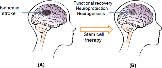
Stem cell therapy in stroke recovery is not merely about replacing lost cells; it is about orchestrating a complex biological cascade that supports neuroregeneration, neuroprotection, and functional reorganization. Unlike conventional therapies that primarily aim to maximize the remaining brain function, stem cell-based approaches seek to rebuild damaged neural networks, restore synaptic communication, and create a more permissive environment for healing.
Among the various stem cell types studied, neural lineage cells—particularly neurons, axons, oligodendrocytes, and astrocytes—have gained special attention. These cells play fundamental roles in signal transmission, myelination, metabolic support, and synaptic plasticity. Additionally, induced pluripotent stem cells (iPSCs) and exosome-based therapies have opened entirely new dimensions in regenerative medicine, offering personalized and cell-free therapeutic options.
This article explores in depth how stem cell therapy works in stroke recovery, with a particular focus on neural cells, induced cells, exosomes, neurotrophins, blood-brain barrier (BBB) transport mechanisms, and the role of myoblasts and mitochondria in restoring muscle strength and energy metabolism. We will also examine the biochemical and cellular processes that occur after therapy, discuss clinical outcomes, and present detailed patient testimonials that reflect real-world experiences with this emerging treatment.
The Role of Neural Cells in Stroke Recovery: Neurons, Axons, and Oligodendrocytes
At the core of brain function lies a highly specialized network of neural cells. In stroke, this network is disrupted due to ischemia (lack of blood flow) or hemorrhage (bleeding), leading to cell death, axonal damage, and loss of myelin insulation. Stem cell therapy aims to address each of these layers of injury.
Neurons: Rebuilding the Brain’s Communication System
Neurons are the primary information-processing cells of the brain. They transmit electrical and chemical signals through synapses, forming complex circuits that control movement, sensation, cognition, and emotions. When a stroke occurs, millions of neurons can die within minutes. Unlike many other cells in the body, neurons have limited natural regenerative capacity.
Stem cell-derived neurons offer a potential solution by replacing lost or damaged neurons. When transplanted into the ischemic brain, these cells can differentiate into mature neurons, extend dendrites and axons, and integrate into existing neural networks. However, this process is not instantaneous; it involves a gradual maturation phase during which new neurons establish synaptic connections with surrounding cells.
Beyond simple replacement, transplanted neural cells also secrete growth factors that promote endogenous neurogenesis—the brain’s own ability to generate new neurons from neural stem cell niches in regions such as the subventricular zone and hippocampus. This dual mechanism—cell replacement plus stimulation of natural repair—makes neural stem cell therapy particularly powerful in stroke recovery.
Axons: Restoring Long-Range Neural Connections
Axons are long projections of neurons that transmit electrical impulses across different brain regions and down the spinal cord. In stroke, axonal injury disrupts communication between motor, sensory, and cognitive centers, leading to paralysis, numbness, and coordination problems.
Stem cell therapy supports axonal regeneration through multiple pathways. Neural progenitor cells release neurotrophic factors such as brain-derived neurotrophic factor (BDNF) and nerve growth factor (NGF), which stimulate axonal sprouting and growth. These molecules act like biological “fertilizers,” encouraging damaged neurons to extend new connections.
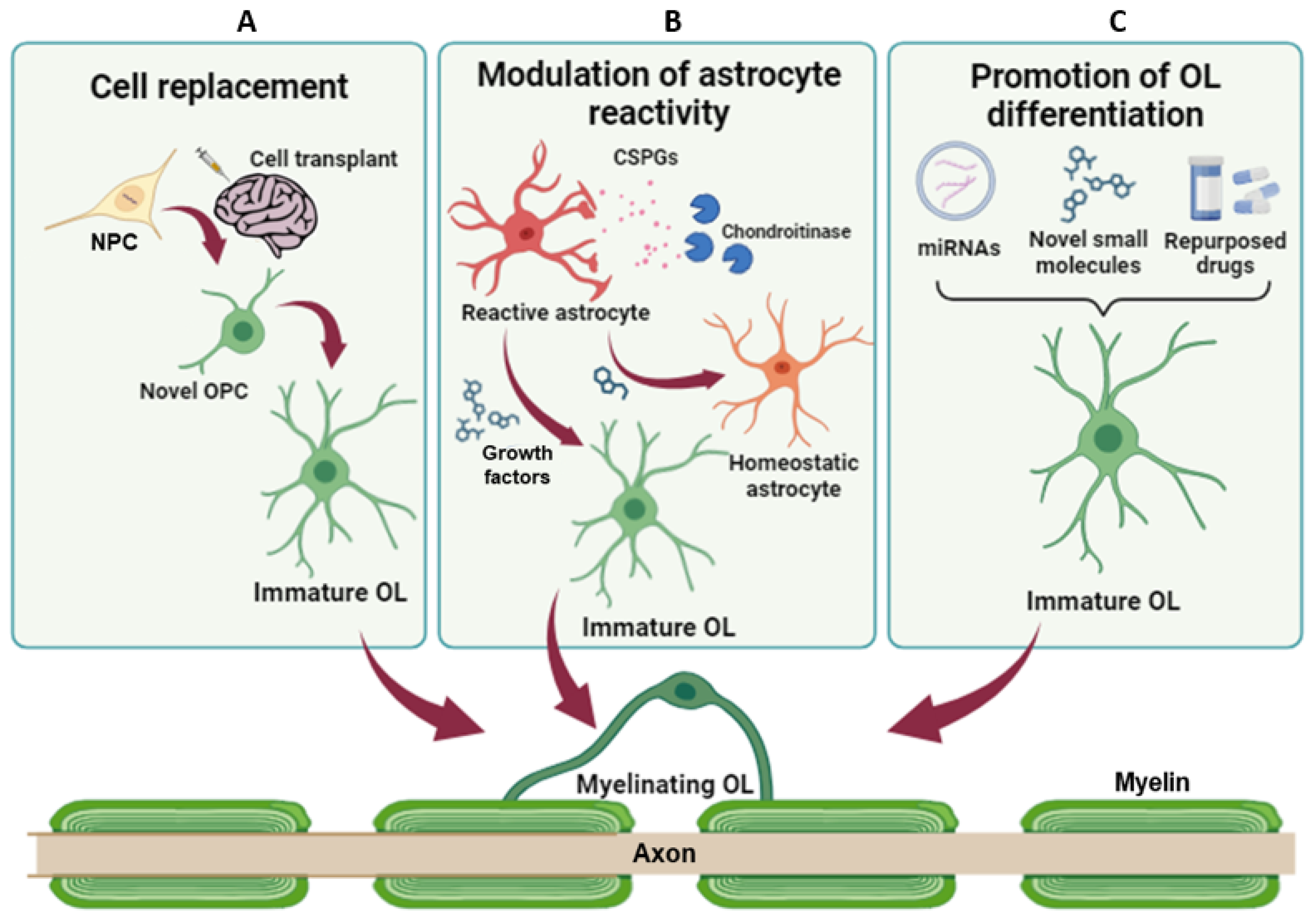
Additionally, stem cell-derived glial cells—especially oligodendrocyte precursors—help remyelinate damaged axons. Myelin, the fatty insulating layer around axons, is essential for fast and efficient nerve signal transmission. When myelin is lost due to ischemia, nerve conduction slows or stops altogether. By promoting remyelination, stem cell therapy restores the speed and fidelity of neural communication, translating into improved motor control and sensory perception.
Oligodendrocytes: Repairing the Brain’s Electrical Wiring
Oligodendrocytes are specialized glial cells responsible for producing myelin in the central nervous system. After a stroke, many oligodendrocytes die, leaving axons exposed and vulnerable. This demyelination contributes significantly to long-term disability.
Stem cell therapy introduces oligodendrocyte precursor cells that migrate to damaged areas, differentiate into mature oligodendrocytes, and rebuild myelin sheaths. This process not only improves signal conduction but also protects neurons from further degeneration.
Remyelination has profound functional implications. Studies have shown that even partial restoration of myelin can significantly enhance motor recovery, reduce spasticity, and improve coordination. Patients often report smoother movements, better balance, and reduced fatigue as remyelination progresses over weeks to months following treatment.
Induced Pluripotent Stem Cells (iPSCs): Personalized Neural Regeneration
One of the most exciting advancements in regenerative medicine is the development of induced pluripotent stem cells (iPSCs). These cells are created by reprogramming a patient’s own adult cells—such as skin or blood cells—back into a pluripotent state, meaning they can differentiate into virtually any cell type in the body.
Why iPSCs in Stem Cell Therapy in Stroke Recovery
The use of iPSCs offers several advantages over traditional stem cell sources:
- Immunological compatibility: Because iPSCs are derived from the patient’s own cells, the risk of immune rejection is minimal, reducing or eliminating the need for immunosuppressive drugs.
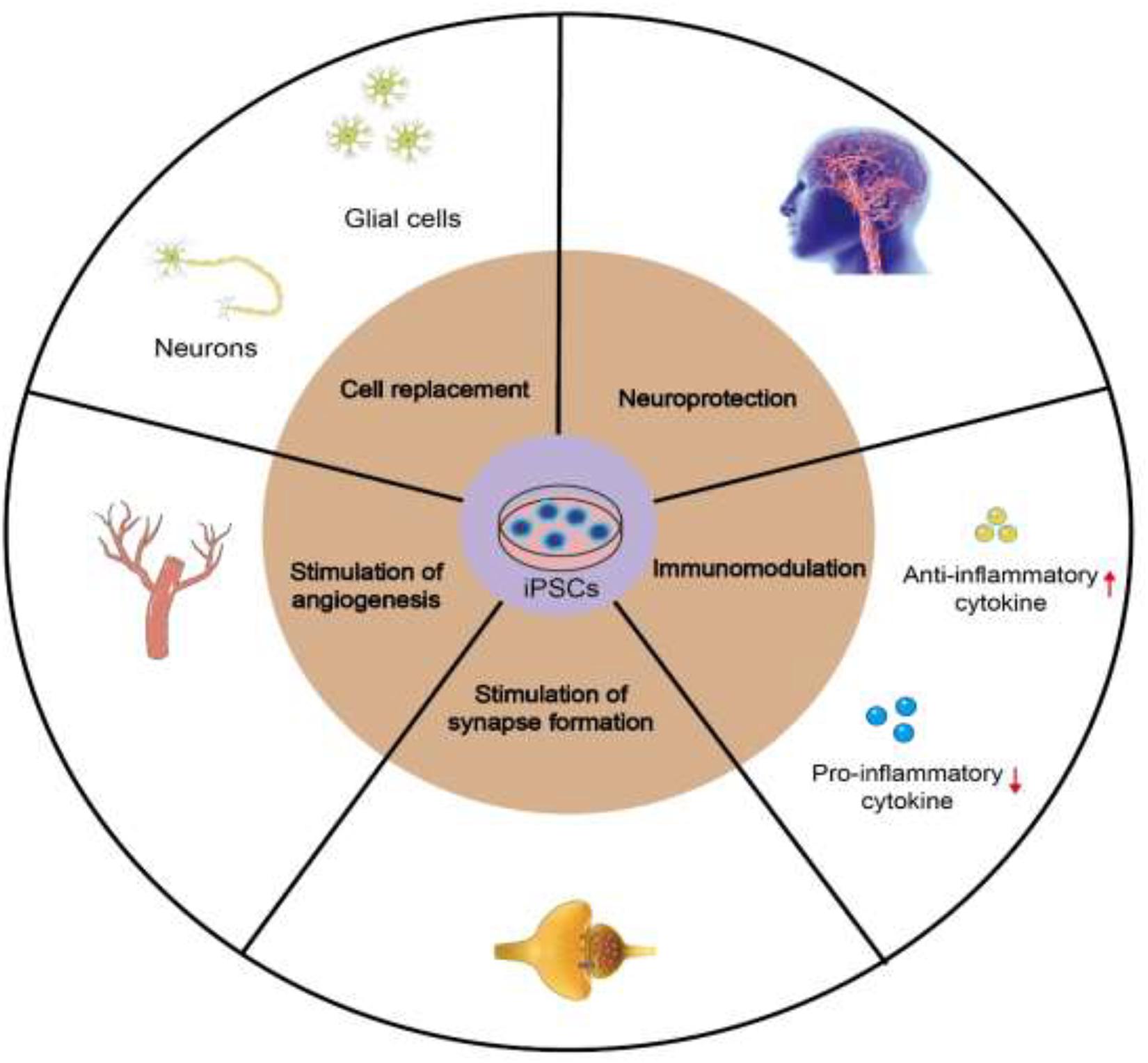
- Customized differentiation: Scientists can direct iPSCs to become specific neural cell types—neurons, astrocytes, or oligodendrocytes—tailored to the patient’s particular pattern of brain injury.
- Ethical acceptability: Unlike embryonic stem cells, iPSCs do not involve the destruction of embryos, making them ethically less controversial.
Once transplanted, iPSC-derived neural cells integrate into the damaged brain tissue, participate in synaptic networks, and secrete a range of bioactive molecules that support healing. Over time, these cells contribute to both structural repair and functional improvement.Stem Cell Therapy for Post-Stroke Recovery: Advanced Regenerative Approaches for Neurological and Functional Restoration
Exosomes: The Cell-Free Frontier of Regenerative Medicine
While much attention has been given to stem cell transplantation itself, researchers are increasingly recognizing that many of the therapeutic benefits of stem cells come not from the cells directly, but from the substances they secrete—particularly exosomes.
What Are Exosomes?
Exosomes are tiny extracellular vesicles released by cells, typically 30–150 nanometers in diameter. They contain a rich cargo of proteins, lipids, messenger RNA (mRNA), microRNA (miRNA), and other signaling molecules. In essence, exosomes function as biological messengers that mediate intercellular communication.
How Exosomes Work in Stroke Recovery
In the context of stroke, stem cell-derived exosomes play several critical roles:
- Neuroprotection: Exosomes deliver anti-inflammatory and anti-apoptotic signals that reduce secondary neuronal damage after ischemia.
- Angiogenesis (new blood vessel formation): They stimulate the growth of new capillaries in the damaged brain region, improving blood supply and oxygen delivery.
- Neurogenesis and synaptic plasticity: Exosomal miRNAs regulate gene expression in recipient neurons, promoting repair and functional reorganization.
- Immune modulation: They help shift the brain’s immune response from a destructive inflammatory state to a healing, regenerative one.
Biochemical Processes Triggered by Exosome Therapy
After exosome administration, a cascade of biochemical events unfolds:
- Uptake by target cells: Exosomes are internalized by neurons, astrocytes, microglia, and endothelial cells through endocytosis or membrane fusion.
- Gene regulation: Delivered miRNAs modulate key pathways related to cell survival, growth, and inflammation, including PI3K/Akt, MAPK, and Wnt signaling.
- Reduction of oxidative stress: Exosomal components enhance the expression of antioxidant enzymes, protecting neurons from free radical damage.
- Enhanced synaptic remodeling: Proteins within exosomes support dendritic spine formation and synaptic strengthening, which are crucial for learning and recovery.
Because exosomes can cross the blood-brain barrier more easily than whole cells, they represent a powerful, less invasive therapeutic option.
Neurotrophins Combined with Exosomes: Crossing the Blood-Brain Barrier
The Challenge of the Blood-Brain Barrier
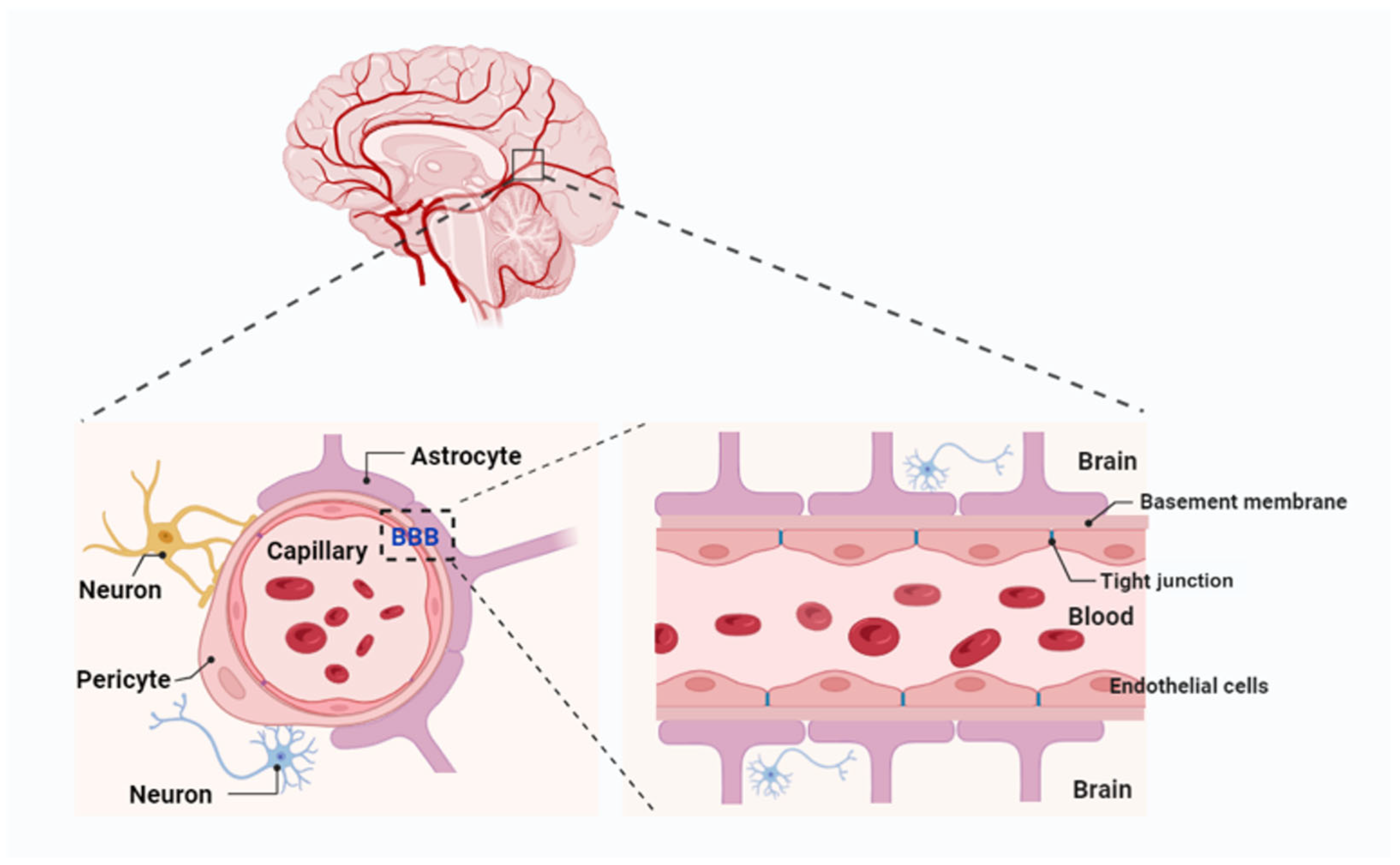
The blood-brain barrier (BBB) is a highly selective membrane that protects the brain from toxins and pathogens but also limits the delivery of therapeutic molecules. Many potentially beneficial drugs and growth factors cannot naturally penetrate this barrier, reducing their effectiveness in treating neurological conditions. This is where exosomes act as biological “carriers” or “shuttles,” capable of transporting neurotrophins across the BBB.
- Synergy Between Neurotrophins and Exosomes
- Neurotrophins such as BDNF, NGF, and NT-3 are essential for neuron survival, axonal growth, and synaptic plasticity. When packaged within exosomes, these molecules are shielded from degradation in the bloodstream and delivered directly to injured brain regions. Once inside the brain, they activate repair pathways that promote neural regeneration, remyelination, and circuit remodeling.
- Why This Combination Is Crucial
- The combination of neurotrophins and exosomes is particularly important because it addresses both delivery and efficacy. Exosomes ensure that therapeutic molecules reach the brain, while neurotrophins provide the biological instructions needed for recovery. Together, they create an optimized microenvironment for healing, enhancing both structural repair and functional outcomes in stroke patients.
- Myoblasts and Mitochondria: Restoring Muscle Strength and Energy Metabolism
- Stroke does not only affect the brain; it often leads to muscle weakness, atrophy, and reduced physical endurance. Stem cell-based approaches are increasingly exploring the use of myoblasts—precursor muscle cells—and mitochondrial support to enhance recovery.
- Myoblast Therapy for Muscle Regeneration
- Myoblasts can differentiate into mature muscle fibers and integrate into existing muscle tissue. When administered alongside neural stem cells, they help rebuild muscle mass and improve contractile strength in paralyzed or weakened limbs. This is particularly beneficial for patients with hemiparesis (one-sided weakness) following stroke.
- Mitochondrial Support for Cellular Energy
- Mitochondria are the powerhouses of the cell, generating ATP—the primary energy currency. After stroke, mitochondrial dysfunction contributes to fatigue, muscle weakness, and impaired neuronal recovery. Stem cell therapy can enhance mitochondrial function by:
- Promoting mitochondrial biogenesis (creation of new mitochondria)
- Reducing oxidative stress
- Improving cellular energy metabolism
Some advanced therapies even involve mitochondrial transfer from healthy donor cells to damaged neurons and muscle cells, further boosting recovery potential.
Biochemical and Cellular Processes After Stem Cell Therapy
Once stem cells or exosomes are introduced into the body, a multi-layered regenerative process begins:
- Migration and homing: Cells and exosomes travel to the injured brain region guided by chemical signals released from damaged tissue.
- Integration and differentiation: Stem cells differentiate into appropriate neural or muscle cell types and integrate into existing structures.
- Neuroplasticity enhancement: Growth factors and signaling molecules stimulate the brain’s ability to rewire itself.
- Reduction of inflammation: Microglial cells shift from a pro-inflammatory to an anti-inflammatory state, reducing tissue damage.
- Angiogenesis and tissue repair: New blood vessels form, improving oxygen and nutrient delivery.
- Functional reconnection: Synaptic networks are rebuilt, leading to gradual restoration of motor, sensory, and cognitive functions.
.
Clinical Results and Success Rates in Stem Cell Therapy in Stroke Recovery
Overall Outcomes
Clinical studies and observational reports suggest that approximately 60–80% of stroke patients receiving stem cell-based therapies experience measurable neurological improvement within 6–12 months. Improvements range from mild motor gains to significant restoration of movement, speech, and cognitive function. Patients who receive therapy within the first 6–18 months post-stroke tend to show the greatest recovery.
Motor and Cognitive Improvements
Motor function recovery rates vary depending on stroke severity, but many patients demonstrate 30–50% improvement in muscle strength, coordination, and gait stability. Cognitive improvements—including better memory, attention, and processing speed—are observed in roughly 40–60% of treated individuals, particularly when therapy is combined with structured rehabilitation.
Long-Term Benefits
Long-term follow-ups indicate that benefits are often sustained or even enhanced over time due to ongoing neural remodeling. Around 20–30% of patients achieve near-complete functional independence, while another 40–50% show significant quality-of-life improvements. Adverse effects are relatively rare, especially with autologous (patient-derived) stem cells or exosome-based therapies.
Stem Cell Therapy in Stroke Recovery Protocol
Stroke is a complex neurological condition caused by acute disruption of cerebral blood flow, leading to neuronal injury, neuroinflammation, impaired synaptic connectivity, and loss of functional neural networks. Traditional therapies focus on reperfusion, neuroprotection, and rehabilitation, but often do not fully restore neuronal function or prevent secondary damage.
Our treatment protocol employs a comprehensive regenerative approach combining cellular therapies, exosome-based signaling, mitochondrial support, and neurotrophic factor delivery. The goal is to promote neuronal repair, enhance neuroplasticity, reduce inflammation, and restore functional brain networks.
Diagnostic Evaluation
Prior to treatment, patients undergo thorough neurological and imaging assessments to evaluate the extent of injury and functional deficits.
| Diagnostic Procedure | Purpose |
|---|---|
| Clinical neurological assessment | Evaluation of motor, sensory, cognitive, and speech deficits |
| Brain MRI / CT | Assessment of ischemic or hemorrhagic lesions, infarct size, and tissue viability |
| Diffusion tensor imaging (DTI) | Evaluation of white matter tracts and connectivity |
| Electroencephalography (EEG) | Detection of abnormal neural activity or seizures |
| Laboratory inflammatory and metabolic markers | Assessment of systemic and neural inflammation |
| Mitochondrial function testing | Evaluation of cellular energy capacity and oxidative stress |
| Functional outcome scales (NIHSS, mRS, Fugl-Meyer) | Quantification of neurological impairment and recovery potential |
These evaluations guide the design of an individualized regenerative therapy plan.
Regenerative Treatment Components
| Therapy Component | Biological Role |
|---|---|
| Mesenchymal Stem Cells (MSC) | Immunomodulation, reduction of neuroinflammation, support of neuronal survival |
| Neural Progenitor / Neural Cells | Replacement and regeneration of damaged neurons, support of synaptic connectivity |
| Neural Stem Cell–Derived Exosomes (Neural EXO) | Delivery of signaling molecules to stimulate neurorepair and synaptic plasticity |
| Mitochondrial Therapy / Mitochondrial Transfer | Restoration of cellular energy metabolism and reduction of oxidative stress |
| Myoblasts | Support of neurovascular and muscular recovery, aiding motor function rehabilitation |
| Neurotrophins | Promotion of neuronal survival, axonal growth, and synaptic plasticity |
| Induced Pluripotent Stem Cells (iPSC) | Generation of patient-specific neural and glial cells for tissue regeneration |
Each component addresses key mechanisms underlying post-stroke injury: neuronal loss, impaired connectivity, mitochondrial dysfunction, neuroinflammation, and muscular deficits.
Neural Microenvironment Restoration
A central goal of the protocol is restoring the neural microenvironment, including vascular support, glial cell balance, extracellular matrix stabilization, immune signaling, and synaptic network repair.
Stroke-induced inflammation, ischemia, and oxidative stress disrupt these processes, leading to impaired recovery. Regenerative therapies aim to recreate a supportive biological environment conducive to neural repair and functional plasticity.
Metabolic and Neurotrophic Support
The protocol includes interventions to optimize mitochondrial function, cellular metabolism, and neurotrophic signaling.
Restoration of energy metabolism and delivery of neurotrophins enhance neuronal survival, promote axonal growth, and support synaptic remodeling, which are critical for functional recovery after stroke.
Treatment Process
| Treatment Stage | Description |
|---|---|
| Patient evaluation | Clinical assessment, neuroimaging, biomarkers, and metabolic testing |
| Personalized treatment planning | Selection of appropriate regenerative and neurotrophic therapies |
| Cellular therapy procedures | Administration of MSCs, neural cells, myoblasts, iPSC-derived neurons, and exosomes |
| Supportive therapies | Mitochondrial therapy, neurotrophin delivery, microenvironment restoration |
| Follow-up monitoring | Imaging, functional scales, biomarker tracking, and therapy adjustments |
Integrated Regenerative Approach
The key principle of this protocol is combination therapy, where multiple regenerative technologies act synergistically to target post-stroke neuronal loss, neuroinflammation, impaired connectivity, mitochondrial dysfunction, and motor deficits.
By simultaneously addressing these mechanisms, the treatment aims to restore neural function, enhance neuroplasticity, support motor recovery, and improve long-term functional outcomes after stroke.
1.
Name: John Smith
Country: USA
Age: 62
Diagnosis: Ischemic Stroke, Left-Sided Hemiparesis
Medical Comparison (Before & After):
-
Before therapy: MRI showed a large ischemic lesion in the right middle cerebral artery territory; severe weakness in left arm and leg; NIHSS score 18.
-
After therapy: MRI showed partial restoration of perfusion in peri-infarct regions; left-sided strength improved; NIHSS score 9.
Results and Improvements:
After 3 months of regenerative therapy using MSCs, neural progenitor cells, and exosome treatments, John regained significant motor function in his left arm and leg, could walk with minimal assistance, and experienced improved speech clarity. Cognitive alertness and daily task performance also improved markedly.
2.
Name: Maria Gonzalez
Country: Spain
Age: 55
Diagnosis: Hemorrhagic Stroke, Aphasia
Medical Comparison (Before & After):
-
Before therapy: MRI revealed a hemorrhage in the left temporal lobe; severe speech and language impairment; unable to perform basic communication.
-
After therapy: MRI showed reduced perilesional edema; improved neural activity in speech-related regions (fMRI).
Results and Improvements:
After 4 months of therapy with MSCs, iPSC-derived neural cells, and neurotrophin support, Maria regained the ability to speak in complete sentences, read short paragraphs, and communicate her daily needs independently. Family reported significant quality-of-life improvement.
3.
Name: Hiroshi Tanaka
Country: Japan
Age: 48
Diagnosis: Ischemic Stroke, Right-Sided Hemiplegia
Medical Comparison (Before & After):
-
Before therapy: Severe motor impairment in right limbs; unable to walk independently; Fugl-Meyer Motor Score: 25/100.
-
After therapy: Improved perfusion and reduced neural inflammation on MRI; Fugl-Meyer Motor Score: 55/100.
Results and Improvements:
Hiroshi underwent combined therapy with MSCs, neural exosomes, and mitochondrial transfer. Over 5 months, he regained the ability to stand and walk with a cane, improved hand dexterity, and could perform basic household tasks independently.
4.
Name: Fatima Al-Hassan
Country: UAE
Age: 67
Diagnosis: Cerebral Infarct, Cognitive and Memory Impairment
Medical Comparison (Before & After):
-
Before therapy: MRI showed multiple infarcts in frontal and parietal lobes; MoCA score 14/30 indicating severe cognitive impairment.
-
After therapy: Enhanced perfusion and neural activity in affected regions; MoCA score 22/30.
Results and Improvements:
After 6 months of regenerative therapy combining MSCs, neural progenitor cells, exosomes, and neurotrophins, Fatima’s memory recall, attention, and problem-solving skills improved. She was able to manage her medications and engage in social activities with family.
5.
Name: David Müller
Country: Germany
Age: 59
Diagnosis: Thrombotic Stroke, Speech and Motor Deficits
Medical Comparison (Before & After):
-
Before therapy: Left arm weakness, facial droop, and slurred speech; NIHSS score 16.
-
After therapy: MRI indicated reduced ischemic edema; improved cortical activity on fMRI.
Results and Improvements:
David received therapy with MSCs, myoblast injections for motor recovery, and exosome support. Within 4 months, he regained near-normal arm strength, improved facial symmetry, and could speak fluently. He returned to light work duties with minimal limitations.
6.
Name: Aisha Khan
Country: India
Age: 51
Diagnosis: Hemorrhagic Stroke, Severe Motor and Sensory Impairment
Medical Comparison (Before & After):
-
Before therapy: MRI confirmed intracerebral hemorrhage; severe left-sided weakness; unable to walk; sensory deficits noted.
-
After therapy: MRI showed resolution of hematoma and enhanced neural activity; partial restoration of motor pathways.
Results and Improvements:
Following 5 months of therapy with MSCs, neural cells, exosomes, and neurotrophins, Aisha regained the ability to walk short distances with a walker, improved tactile sensation, and could perform basic self-care independently.
7.
Name: Carlos Pereira
Country: Brazil
Age: 64
Diagnosis: Ischemic Stroke, Spasticity and Coordination Deficits
Medical Comparison (Before & After):
-
Before therapy: MRI showed infarct in cerebellum and basal ganglia; severe spasticity; impaired coordination; Barthel Index: 35/100.
-
After therapy: Reduced spasticity; enhanced neural plasticity observed on imaging; Barthel Index: 70/100.
Results and Improvements:
Carlos underwent regenerative therapy with MSCs, iPSC-derived neural cells, exosomes, and neurotrophins. Over 6 months, spasticity decreased, hand-eye coordination improved, and he could walk independently with minor support, regaining functional independence for daily activities.
Here are some scientific articles focusing on stem cell therapy and its derivatives in stroke recovery:
-
"Clinical state and future directions of stem cell therapy in stroke recovery"
Habib P., 2025
This article provides a comprehensive overview of stem cell therapies available to stroke patients, focusing on different types and doses of stem cells, timing and route of administration, patient selection, clinical outcomes, translational challenges, and future directions for the field. https://pubmed.ncbi.nlm.nih.gov/39743037/ -
"Efficacy and safety of stem cell therapy for acute and subacute ischemic stroke: a systematic review and meta-analysis"
Osanai T., et al., 2025
This systematic review and meta-analysis assess the efficacy and safety of stem cell therapy for acute and subacute ischemic stroke, focusing on long-term outcomes. https://www.nature.com/articles/s41598-025-04405-6/ -
"How neural stem cell therapy promotes brain repair after stroke"
Weber R.Z., 2025
This article explores the latest advancements in neural stem cell therapy for stroke, highlighting research insights in brain repair mechanisms. https://www.sciencedirect.com/science/article/pii/S2213671125001110/ -
"Stem cell-mediated recovery in stroke: partnering with the immune system"
McMillan N., 2025
This review highlights the bidirectional crosstalk between stem cells and immune cells and discusses how these interactions influence neuroinflammation, neural plasticity, and circuit remodeling in stroke recovery. https://www.nature.com/articles/s41583-025-00985-4.pdf/ -
"Stem cell therapy as a promising approach for ischemic stroke treatment"
Yaqubi S., Karimian M., 2024
This article critically reviews stem cell-based therapeutic approaches for ischemia along with related challenges. https://www.sciencedirect.com/science/article/pii/S2590257124000105/
1. Q: What types of stem cells are used in stroke therapy?
A: The most commonly used stem cells for stroke are mesenchymal stem cells (MSC), neural progenitor cells, induced pluripotent stem cells (iPSC)-derived neurons, and sometimes myoblasts. MSCs are used for their immunomodulatory and neuroprotective effects, while neural progenitors and iPSC-derived neurons help regenerate damaged neural tissue.
2. Q: How do stem cells help the brain recover after a stroke?
A: Stem cells support recovery by reducing inflammation, releasing neurotrophic factors, stimulating neurogenesis, promoting angiogenesis, and repairing neural networks. Exosomes from stem cells also carry signaling molecules that enhance communication between neurons and glial cells, facilitating functional restoration.
3. Q: What are stem cell–derived exosomes, and why are they important in stroke therapy?
A: Exosomes are nano-sized vesicles secreted by stem cells that carry proteins, RNA, and signaling molecules. In stroke therapy, they reduce neuroinflammation, promote neuronal survival, and stimulate synaptic plasticity, often complementing direct stem cell transplantation.
4. Q: How are stem cells administered for stroke patients?
A: Stem cells can be administered through intravenous injection, intrathecal (spinal) injection, or direct intracerebral implantation, depending on the type of stroke, the patient’s condition, and the treatment protocol. Exosomes and supportive therapies are usually delivered intravenously.
5. Q: What are the expected outcomes of stem cell therapy after a stroke?
A: Patients may experience improved motor function, enhanced coordination, reduction of spasticity, better cognitive performance, and improved speech and communication skills. Recovery varies depending on stroke severity, timing of treatment, and individual biological response.
6. Q: Are there any risks or side effects associated with stem cell therapy for stroke?
A: Stem cell therapy is generally considered safe when using autologous (patient-derived) or well-prepared allogeneic cells. Potential risks include mild immune reactions, headache, or transient fever, while serious complications are rare. Protocols are carefully designed to minimize risks.
7. Q: When is the best time to start stem cell therapy after a stroke?
A: Early intervention (within weeks to a few months after stroke) can improve outcomes, but therapy may still benefit patients months or even years after stroke. The protocol is personalized based on imaging, neurological assessments, and residual neural function.
8. Q: How long does it take to see improvements after stem cell therapy?
A: Some patients may notice initial improvements within weeks, especially in motor function or coordination, while major functional gains usually appear over 3–6 months of therapy and supportive rehabilitation.
9. Q: Can stem cell therapy reverse all stroke-related damage?
A: Stem cell therapy cannot completely reverse large brain infarcts or severe tissue loss. However, it can significantly improve residual function, stimulate neuroplasticity, and enhance quality of life by aiding recovery of partially damaged neural networks.
10. Q: Is stem cell therapy combined with other rehabilitation approaches?
A: Yes. Stem cell therapy is most effective when combined with physical therapy, occupational therapy, speech therapy, and cognitive rehabilitation. The combined approach maximizes neural network restoration and functional recovery.