

Fetal hydrocephalus is one of the most complex diseases of childhood. It has many causes, including loss of brain tissue (cerebral atrophy), overproduction of cerebrospinal fluid (CSF), or obstruction of CSF pathways due to abnormal neuro- and gliogenesis. Surgical treatments such as CSF shunting and endoscopic third ventriculostomy (ETV) currently used to treat children with intrauterine hydrocephalus are insufficient. It is estimated that 50% of shunts fail within two years, and 20–50% close within five years; infections are also quite common. In addition, we do not yet know what consequences may occur when cerebrospinal fluid proteins enter a confined space, such as the peritoneum, where a large number of immune system cells are located. There is a high probability that bypass surgery and its consequences generate autoantibodies against specific proteins in the cerebrospinal fluid. If these antibodies, or the cells that produce them, end up in the brain, they can alter neuronal physiology and worsen neurological deficits. Regarding the shunt, we do not know the consequences of creating an opening in the membranous floor of the third ventricle to drain cerebrospinal fluid into the subarachnoid space. Given that the floor of the third ventricle is a highly specialized region for the secretion of regulatory factors that influence the activity of the pituitary gland, the shunt may have an adverse effect on neuroendocrine regulation.It can also divert signaling molecules in the cerebrospinal fluid away from their intended targets.
Changes in the composition of cerebrospinal fluid have profound effects on brain development and function. Thus, hydrocephalus and available treatments (shunt surgery and choroid plexus cauterization) may limit the availability of these beneficial factors to the developing and adult brain, resulting in severe lifelong neurological deficits.

The financial and emotional costs of treatment for patients and their families are high, and additional treatments are needed.
It is now recognized that hydrocephalus occurring in the fetus is more than just a disturbance in the dynamics of the cerebrospinal fluid. It is also a brain disease. Recent studies have shown that hydrocephalus and abnormal neurogenesis share a common history: pathology of neural stem cells (NSCs)/radial glial cells that are located in the ventricular zone (VZ) during pregnancy. In hydrocephalus, intercellular junction proteins accumulate abnormally in the cytoplasm of NSCs and ependymal cells and, depending on the stage of brain development, lead to their detachment from the VZ. Violation of the VZ can also be caused by infections or intracerebral hemorrhage. Only a minority of cases are associated with Mendelian inheritance, with X-linked hydrocephalus being the most common type.
The process of destruction begins in the early stages of embryonic life and ends during the first weeks of postnatal life. At the end of development, some areas are devoid of ependymal and sub ependymal cells, while others are not.
Since the disorder is genetically determined and its consequences are widespread, questions arise: can this process be reversed and will this have a therapeutic effect?
The available data give hope for the possibility of using regenerative therapies based on the use of neural stem cells. These cells have two main characteristics: they are self-sustaining and pluripotent. This means that they reproduce to renew themselves; they also have the potential to differentiate into multiple types of brain cells.
Hydrocephalus is a neurological condition characterized by excessive accumulation of cerebrospinal fluid (CSF) in the ventricular system, leading to increased intracranial pressure, ventricular enlargement, white matter damage, and progressive cognitive and motor impairment. While ventriculo peritoneal shunting remains the gold standard of treatment, regenerative medicine approaches are being investigated to address secondary brain injury, inflammation, and tissue degeneration associated with chronic hydrocephalus.
Neural stem/progenitor cells (NSCs) are among the most biologically relevant cell types for this condition. These cells can differentiate into neurons, astrocytes, and oligodendrocytes. In experimental settings, NSCs may support repair of periventricular white matter, promote remyelination, enhance synaptic plasticity, and improve neuronal survival. They also secrete neurotrophic factors such as BDNF and GDNF, which help reduce apoptosis and support neural network recovery.
Mesenchymal stem cells (MSCs), derived from bone marrow, adipose tissue, or umbilical cord tissue, exert primarily paracrine and immunomodulatory effects. In hydrocephalus models, MSCs have been shown to reduce neuroinflammation, suppress microglial activation, decrease oxidative stress, and protect the blood-brain barrier. Their anti-inflammatory cytokine secretion may limit secondary injury caused by chronic ventricular dilation and intracranial pressure fluctuations.
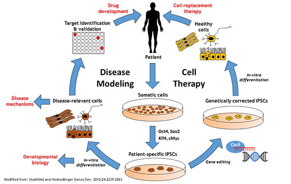
Induced pluripotent stem cell (iPSC)-derived neural cells represent a more advanced strategy. These cells can be directed into specific neuronal or glial lineages, potentially allowing targeted replacement of damaged cell populations. iPSC-derived oligodendrocyte progenitors may help restore myelin integrity in periventricular regions affected by long-standing hydrocephalus.
Exosome-based therapies, derived from MSC or NSC cultures, provide a cell-free alternative. Exosomes contain growth factors, microRNAs, and anti-inflammatory mediators that may enhance neuroprotection, reduce fibrosis, and improve intercellular signaling without the risks associated with live cell transplantation.
Biobanked allogeneic cell cultures offer standardized, quality-controlled therapeutic material, improving safety and reproducibility. Although these approaches remain largely investigational, they aim to complement surgical treatment by reducing inflammation, supporting neural repair, improving white matter integrity, and potentially enhancing cognitive and functional recovery in patients with hydrocephalus.
There are at least five benefits of using neural cells for neural repair strategies:
NSCs are available for cultivation
they can be transplanted
they migrate
differentiate and integrate into damaged areas
In addition, studies have shown that the ability of NSCs to migrate and differentiate into the desired cell type depends on damaged sites that release specific chemotactic factors.
Healthy NSCs are transplanted into the cerebrospinal fluid to replace radial glial cells, neural progenitors, and neuroblasts that are lost during the hydrocephalic process. This regenerative therapy can restore the VZ and/or reverse the effects of VZ destruction. Thus, malformations of the cerebral cortex found in children with hydrocephalus and until now considered incurable have a realistic and promising alternative treatment. Such studies also open up new ways to identify genes and epigenetic factors (growth factors, hormones) involved in the phenotypic differentiation of neurons and glia, which will allow the development of new drugs that regulate normal development of the cerebral cortex.
The development of regenerative therapy based on the use of NSCs for the treatment of children with hydrocephalus is progressing well. Clinical studies have shown that NSCs transplanted into the cerebrospinal fluid migrate to destroyed areas and integrate into neural tissue.
NSCs proliferate to form neurospheres. The NSCs that form these neurospheres express the same cell junction pathology as NSCs.
These pluripotent cells have great clinical potential for tissue repair and also represent a future relief or treatment for a wide range of common disorders. It is hypothesized that replacing a patient’s defective cells with transplantation of hES cell-derived equivalents will restore normal function.
Human brain development is a long process that begins in the third week of pregnancy. Neuronal proliferation begins in the sixth week of pregnancy and is largely completed by mid-gestation. As neurons form, they migrate to different areas of the brain, where they begin to establish connections with other neurons, creating rudimentary neural networks. Although major fiber pathways, including the thalamocortical pathway, are completed at the end of the prenatal period, brain development continues for a long period after birth.
What to expect from treatment?
Stem cells directly replace lost cells, and they can also protect the nervous system through mechanisms other than cell replacement, such as modulation of the immune system. It is worth noting the remarkable ability of NSCs and NPCs to interact with endogenous cells and reconstruct the damaged nervous system when used. In addition, a recent study demonstrated an astrocytic response in the disrupted VZ of hydrocephalus, with astrocytes taking on the morphological and functional characteristics of ependymal cells, suggesting that they function as a CSF-brain barrier involved in the transport of water and solutes. Such remodeling could help restore lost functions at the interface between the brain parenchyma and the cerebrospinal fluid. Thus, we hypothesize that transplantation of NSCs into the hydrocephalic brain will result in cell replacement and/or the creation of a protective microenvironment to prevent progressive destruction of the VZ and enhance beneficial glial responses.
PREPARE AN INDIVIDUAL TREATMENT PLAN
