How Long Do Stem Cells Stay in the Body?
The In-Vivo Life Cycle, Biodistribution, Cellular Communication, and the Role of Extracellular Vesicles Stem cell-based medicine is often described to patien…
The In-Vivo Life Cycle, Biodistribution, Cellular Communication, and the Role of Extracellular Vesicles
Stem cell-based medicine is often described to patients in deceptively simple terms: cells are administered, they travel to the damaged area, repair the tissue, and eventually disappear. The biological reality is considerably more complex. After administration, therapeutic cells enter a dynamic environment in which blood flow, tissue barriers, immune surveillance, cell adhesion, inflammatory signals, extracellular matrix interactions, and intercellular communication determine their fate. In many cell therapies, particularly those based on mesenchymal stromal/stem cells (MSCs), the therapeutic effect may persist even when only a small number of administered cells remain detectable in the target tissue. This observation has shifted the scientific understanding of cell therapy from a simple “replacement of damaged cells” model toward a more complex model involving transient cellular activity, paracrine signaling, immunomodulation, extracellular vesicles (EVs), and transfer of biologically active molecules.
The duration of cell persistence depends strongly on the cell type, source, culture conditions, dose, route of administration, disease state, and methods used to detect the cells. Following intravenous administration, many MSCs are rapidly retained within the pulmonary microcirculation, with subsequent redistribution and clearance involving the liver, spleen, lungs, and immune system. Studies have reported very different persistence times, ranging from hours to days or, depending on the model and detection method, substantially longer periods. Importantly, detection of cellular material does not necessarily mean that living, functionally active cells remain present.Stem Cell Treatment Abroad: An International Clinic with Advanced Regenerative Medicine Solutions
Extracellular vesicles, including populations commonly referred to as exosomes, represent a distinct but closely related therapeutic concept. Rather than introducing whole living cells, cell-free approaches attempt to deliver some of the biologically active signals produced by cells. EVs can transport proteins, lipids, messenger RNA, microRNA, and other molecular components between cells. Their circulation time is generally much shorter than the potential biological duration of their downstream effects, and their biodistribution is influenced by the liver, spleen, lungs, macrophages, vesicle characteristics, and route of administration.
This review examines the complete in-vivo life cycle of therapeutic cells and extracellular vesicles, explains what happens after intravenous and local administration, discusses intracellular and extracellular mechanisms of action, and considers why the question “How long do stem cells stay in the body?” has no single universal answer. For patients, the most important concept is that persistence, biological activity, and therapeutic effect are three different measurements and should not be treated as synonymous.
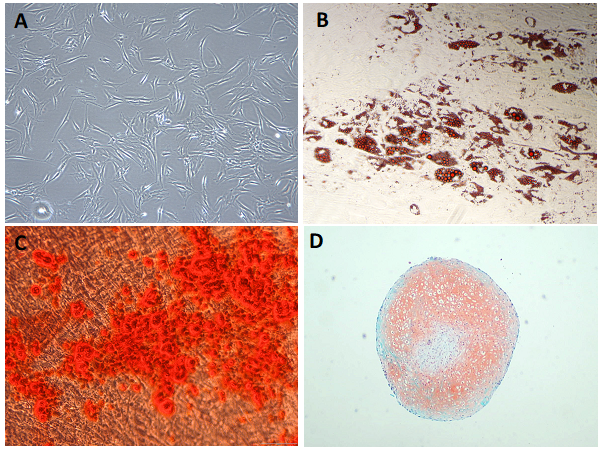
Introduction
One of the most common questions asked about stem cell therapy is also one of the most difficult to answer precisely: How long do the cells remain in the body?
The intuitive assumption is that therapeutic cells behave like transplanted replacement parts. A patient receives cells, the cells travel to a damaged organ, become established there, repair the tissue, and remain permanently. This model may be relevant to some forms of transplantation and cellular replacement, but it does not accurately describe many contemporary regenerative cell therapies.
In particular, mesenchymal stromal cells are increasingly understood not simply as cells that must permanently engraft and transform into new tissue, but as biologically active participants in a temporary signaling process. Their effects may involve interactions with immune cells, endothelial cells, fibroblasts, epithelial cells, resident progenitor cells, extracellular matrix components, and damaged tissue. A substantial part of this communication can occur through molecules released outside the therapeutic cell, including soluble factors and extracellular vesicles.
This distinction is fundamental because the physical presence of a therapeutic cell and its biological influence are not necessarily the same thing.
A cell can be detectable but no longer metabolically active. Conversely, a cell may disappear relatively quickly while having already triggered a cascade of signals that continues after the original cell has been cleared. Similarly, an extracellular vesicle may circulate for a relatively short period but deliver molecular cargo that changes the behavior of a recipient cell for much longer.
Consequently, the in-vivo fate of a cell therapy should be considered as a sequence rather than a single time point:
administration → circulation or local distribution → tissue interaction → cellular signaling → uptake or activation → migration/retention → functional activity → immune recognition and clearance → resolution of the therapeutic signal.
The duration of each stage can vary substantially.
The therapeutic cell is not an isolated biological unit
Once administered, a therapeutic cell immediately becomes part of the patient's biological environment. It encounters plasma proteins, complement components, circulating immune cells, endothelial surfaces, extracellular matrix proteins, oxygen gradients, mechanical forces, inflammatory mediators, and tissue-specific signaling molecules.
The behavior of the cell therefore depends not only on its intrinsic properties but also on the biological environment into which it is introduced.
This is particularly important for MSC-based therapies. MSCs are relatively large cells compared with many blood cells. Their size and physical properties influence their initial distribution after intravenous infusion. Experimental literature indicates that a substantial proportion of intravenously administered MSCs can become temporarily retained in the pulmonary microcirculation. Reviews have reported that approximately 50–80% of administered MSCs may localize to the lungs during the initial phase after intravenous administration, although the exact percentage depends on the system, cell preparation, dose, and measurement method.
This phenomenon is sometimes described as the pulmonary first-pass effect.
It does not necessarily mean that the lungs are the final therapeutic destination.
Rather, the lungs can function as an initial biological filter. Cells can subsequently be cleared, redistributed, fragmented, phagocytosed, or, in some circumstances, participate in signaling within pulmonary tissue.
This observation immediately challenges a common patient misconception: intravenous administration does not mean that every cell freely circulates through the entire body until it independently “finds” the damaged organ.
The process is more selective and more complicated.
What happens immediately after intravenous administration?
Intravenous administration introduces cells directly into the bloodstream. The first minutes are therefore dominated by circulation, cell–blood interactions, vascular geometry, and the mechanical properties of the administered cells.
For MSCs, pulmonary retention can occur rapidly because the cells are substantially larger than the diameter of some microvascular passages. Their physical size, deformability, adhesion properties, and interactions with the vascular endothelium contribute to their distribution.
This means that the first anatomical destination of a cell does not necessarily represent its final biological target.
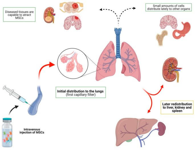
After the initial pulmonary phase, some cellular material may subsequently be detected in organs such as the liver and spleen. Studies have also demonstrated trafficking toward inflammatory or injured tissues.
The degree of redistribution is highly variable.
A patient's inflammatory state can influence the process. Damaged tissues produce chemokines, cytokines, adhesion molecules, and other signals that can alter interactions between circulating cells and the vascular endothelium.
The concept of homing is therefore useful, but it should not be interpreted as a GPS-like mechanism.
Therapeutic cells do not necessarily identify a lesion with perfect precision. Instead, a combination of molecular gradients, adhesion molecules, vascular permeability, tissue inflammation, and local microenvironmental signals can increase the probability that cells or their biological products interact with an injured site.
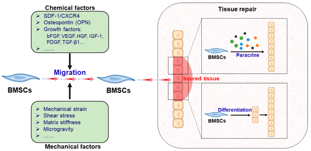
Homing, trafficking, and retention
Cell trafficking involves several interconnected biological processes.
First, cells must remain viable long enough to interact with the circulation and tissue environment. They may then interact with endothelial cells and extracellular matrix components.
Second, the local tissue environment can influence retention.
Inflamed tissues often express higher levels of adhesion molecules and chemotactic signals. These changes can modify the interaction between administered cells and the vascular wall.
Third, after reaching or interacting with the target tissue, the cells can either remain locally, migrate through the tissue, return to circulation, become immobilized, or be removed by immune mechanisms.
The term engraftment should therefore be used carefully.
Detection of a small number of administered cells in a tissue does not automatically demonstrate durable engraftment. In some therapeutic studies, only a very small fraction of the administered cells can be detected at the intended target, even though measurable biological effects occur. Human and animal studies have reported low levels of long-term tissue detection after intravenous MSC administration.
This apparent paradox is one of the central findings of modern cell therapy research.
If relatively few cells remain, how can a therapeutic effect occur?
The answer appears to involve cellular communication.
The paracrine and secretome model
Many therapeutic cells release a complex mixture of biologically active molecules known collectively as the secretome.
The secretome includes soluble proteins, growth factors, cytokines, chemokines, lipids, nucleic acids, and extracellular vesicles.
The therapeutic cell can therefore behave less like a replacement brick and more like a temporary biological communication center.
The cell senses its environment and responds to environmental signals. In turn, it releases molecules that influence surrounding cells.
This creates a dynamic feedback system.
For example, an injured tissue may contain inflammatory mediators that alter the behavior of administered MSCs. The MSCs can then release factors that modify immune-cell activity, endothelial responses, tissue inflammation, apoptosis, and local repair processes.
This mechanism is often called paracrine signaling because the therapeutic cell influences neighboring or nearby cells through released factors.
The process may occur without the therapeutic cell becoming a permanent structural component of the tissue.
This is one reason why disappearance of administered cells does not necessarily mean complete disappearance of their biological influence.
Intracellular communication: what happens inside the recipient cell?
Extracellular communication is only the first step.
The ultimate biological effect often depends on what happens inside the recipient cell.
When a soluble signaling molecule binds to a receptor on a recipient cell, it can activate intracellular signaling pathways. These pathways may modify gene transcription, protein phosphorylation, metabolism, cytoskeletal organization, cell survival, inflammatory signaling, or differentiation programs.
Extracellular vesicles provide another route.
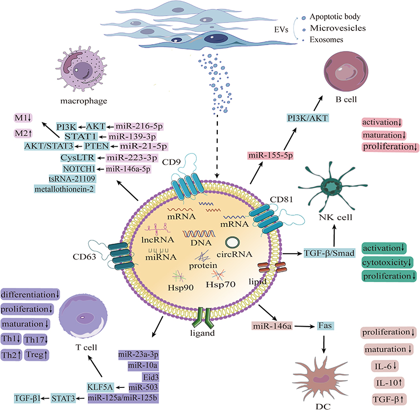
An EV can interact with a target cell through several mechanisms, including receptor-mediated interactions, membrane association, endocytosis, or other uptake processes. Once internalized, its molecular cargo may interact with intracellular regulatory systems.
EV cargo can include proteins, lipids, messenger RNA, microRNA, and other molecular components. The exact composition depends on the producing cell, its state, the culture environment, and the methods used to isolate and characterize the vesicles.
This creates an important biological bridge:
therapeutic cell → secretome/EV release → recipient-cell uptake → intracellular signaling → altered cell behavior.
The therapeutic cell therefore does not necessarily have to remain physically present to initiate downstream biological changes.
Stem cells versus extracellular vesicles
It is useful to distinguish three related but different therapeutic concepts.
The first is whole-cell therapy, in which viable cells are administered.
The second is cell-derived secretome therapy, in which soluble and particulate factors released by cultured cells are used.
The third is extracellular-vesicle-based or cell-free therapy, in which EV-enriched preparations are investigated as therapeutic agents.
These approaches should not be treated as interchangeable.
A living cell is capable of sensing its environment and changing its secretory profile. An isolated EV preparation cannot perform the same degree of adaptive behavior.
On the other hand, EVs are much smaller than cells and do not require the recipient to maintain a population of living donor cells. Their biological activity is therefore based on molecular cargo rather than cellular survival.
This distinction has major implications for pharmacokinetics.
A living cell may remain detectable in a tissue for days or longer under certain circumstances, while an injected EV population can redistribute extremely rapidly and undergo clearance from the bloodstream.Why Stem Cell Therapy Didn’t Work ?
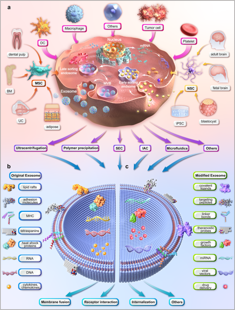
What exactly is an exosome?
The word exosome has become extremely popular in regenerative medicine, but modern scientific terminology is more cautious.
According to the International Society for Extracellular Vesicles, extracellular vesicles are membrane-bound particles released by cells that cannot replicate independently. The term “exosome” should ideally be reserved for vesicles for which an endosomal origin involving multivesicular bodies has been specifically demonstrated. In many experimental preparations, the exact biogenesis of the isolated vesicles cannot be established, so the broader term extracellular vesicles is scientifically more appropriate.
This distinction matters clinically.
A preparation described commercially as “exosomes” may contain a heterogeneous population of extracellular vesicles and other extracellular particles. The composition depends heavily on the cell source, culture conditions, purification method, storage conditions, and characterization techniques.
Therefore, two products carrying the same marketing label may not be biologically equivalent.
The importance of cell culture conditions
A therapeutic cell does not produce exactly the same secretome under every laboratory condition.
Cells respond to their environment.
Oxygen concentration, nutrient availability, cell density, substrate, inflammatory stimulation, passage number, culture medium, serum exposure, and other parameters can influence the molecular composition of the secretome and extracellular vesicles.
This is particularly important for cell-free products.
If two laboratories culture MSCs under different conditions, the resulting EV populations may differ in protein composition, RNA cargo, lipid composition, particle concentration, and biological activity.
The same principle applies to the cells themselves.
Ex-vivo expansion can change cellular phenotype and biological properties. Culture-expanded cells can also differ in size and surface characteristics from their native counterparts. These properties can influence biodistribution after administration.
For this reason, “stem cells” should not be considered a single standardized biological substance.
The biological identity of the cells matters.
Intravenous administration: systemic distribution
Intravenous administration has an obvious advantage: it provides access to the systemic circulation.
However, systemic access does not mean uniform systemic distribution.
For MSCs, the pulmonary circulation is an important initial barrier. A large proportion of cells can be retained in the lungs, after which the detectable cell population may decline and cellular material can appear in other organs.
The liver and spleen are particularly important organs in the subsequent handling of cellular material because of their extensive vascular networks and resident phagocytic cells.
Macrophages and other components of the mononuclear phagocyte system can recognize and remove cellular material.
The therapeutic consequence is fascinating: immune clearance may simultaneously contribute to both the disappearance of the administered cells and the biological response to those cells.
In other words, a therapeutic cell does not necessarily need to survive indefinitely to have an effect.
Its interaction with the immune system may itself become part of the therapeutic mechanism.
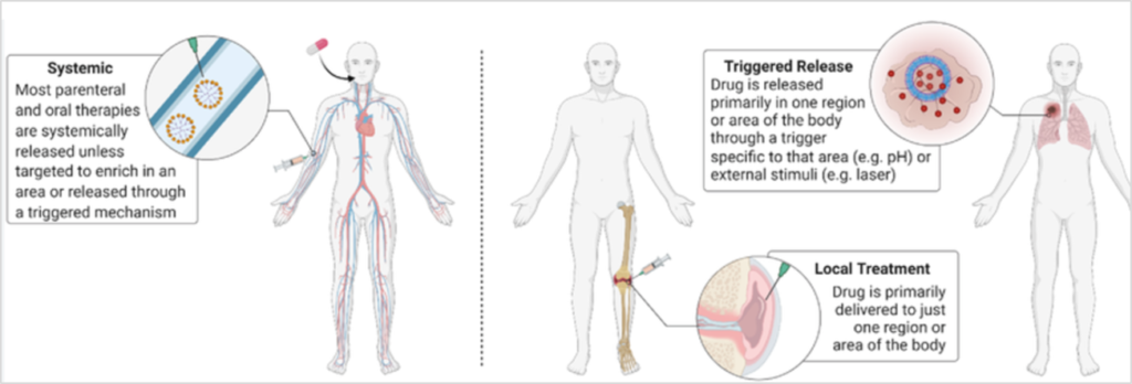
Local administration: a different biological journey
Local administration changes the problem completely.
When cells are injected directly into or near a target tissue, the initial pulmonary first-pass effect associated with intravenous delivery is largely avoided.
The cells begin their biological journey in the vicinity of the intended target.
This can increase local exposure, although it does not guarantee long-term retention.
Local tissue injection can create a highly concentrated cellular microenvironment. However, the injected cells may encounter mechanical stress, local inflammation, extracellular matrix barriers, limited oxygen availability, immune recognition, and other factors that affect survival.
Some cells may remain close to the injection site.
Others may migrate through the tissue or enter local lymphatic or vascular pathways.
Still others may undergo apoptosis or be cleared by resident immune cells.
Thus, local administration may improve anatomical targeting while simultaneously creating a different set of biological challenges.
The optimal route therefore depends on the therapeutic objective rather than on a universal principle that one route is always superior.
The life cycle of an administered cell
The in-vivo life cycle of a therapeutic cell can be conceptualized as a series of overlapping phases.
Immediately after administration, the cell experiences the physical environment of the delivery route.
During the early distribution phase, vascular and tissue interactions determine where the cell is retained.
During the signaling phase, the cell interacts with surrounding cells and releases biological mediators.
During the response phase, the recipient tissue may change its inflammatory, metabolic, vascular, or regenerative behavior.
During the clearance phase, administered cells can undergo apoptosis, necrosis, senescence, phagocytosis, or other forms of elimination.
Importantly, these phases are not strictly sequential.
A cell can simultaneously migrate, secrete molecules, interact with immune cells, and undergo stress responses.
The biological lifetime of the therapeutic effect can therefore exceed the physical lifetime of the administered cell.
How long do stem cells actually stay in the body?
There is no scientifically valid universal number.
Depending on the cell type, administration route, animal model or human study, disease state, dose, cell preparation, labeling strategy, and detection method, persistence can range from hours to days and, in some experimental settings, longer.
For intravenously administered MSCs, some studies have observed rapid disappearance of detectable cells from the blood and lungs. Other studies have reported detectable cellular persistence for substantially longer periods. A review of pulmonary MSC persistence emphasizes that reported duration can vary considerably depending on experimental model and detection method.
This variation is not simply a scientific inconvenience.
It reflects the fact that “cell persistence” is not one measurement.
Researchers can measure:
- intact living cells;
- donor-specific DNA;
- fluorescent labels;
- radiolabels;
- reporter genes;
- donor-derived proteins;
- cell-associated RNA;
- histological signals.
These measurements do not necessarily mean the same thing.
For example, donor DNA can remain detectable after a living donor cell has disappeared. A fluorescent label can remain associated with cellular debris. A reporter signal may disappear because gene expression changes rather than because the cell has died.
Therefore, the statement “the cells are still present” requires clarification: what exactly was detected, and does that detection demonstrate viable and functional cells?
The difference between persistence and therapeutic activity
This is perhaps the most important concept for patients.
Imagine that administered MSCs remain detectable for only a relatively short period.
That does not automatically mean that the treatment was ineffective.
The cells may have released signaling molecules during that period. Those molecules may have altered immune-cell behavior, endothelial responses, local inflammatory pathways, or tissue-resident cells.
The therapeutic response may therefore continue after the administered cells are no longer detectable.
Conversely, detecting donor cells months later would not automatically prove that they remain therapeutically active.
Thus:
cell persistence ≠ cell activity ≠ therapeutic effect.
These are three separate biological variables.
The life cycle of extracellular vesicles
Extracellular vesicles have a different in-vivo life cycle from whole cells.
After systemic administration, EVs are small enough to circulate through vascular compartments that cannot accommodate whole cells.
However, this does not mean that they remain in circulation for a long time.
Experimental biodistribution studies generally demonstrate rapid distribution and clearance of intravenously administered EVs. A substantial proportion may be removed from the bloodstream quickly, with accumulation particularly associated with organs such as the liver and spleen.
The liver is especially important because resident macrophages, including Kupffer cells, can take up extracellular vesicles.
The spleen is another major site of EV interaction with immune cells.
Depending on their physicochemical properties, EVs may also reach the lungs, kidneys, gastrointestinal tissues, and other organs.
Their biodistribution is influenced by vesicle size, surface composition, source cell, route of administration, and disease state.
Why EV circulation can be short but biological effects longer
A short plasma residence time does not necessarily mean a short biological effect.
Consider a simplified example.
An EV enters the bloodstream and is rapidly taken up by a recipient cell. The EV itself may no longer be detectable in plasma, but its cargo can alter intracellular signaling.
If the cargo changes gene expression or protein activity, the recipient cell can remain functionally altered after the original EV has disappeared.
This creates another important distinction:
EV pharmacokinetics and EV pharmacodynamics are not identical.
Pharmacokinetics describes where the particles go and how quickly they disappear.
Pharmacodynamics concerns what biological effect they produce.
The second may outlast the first.
Extracellular vesicles as biological messengers
EVs can be considered a form of intercellular communication system.
Cells release vesicles containing molecular information derived from their internal state.
A recipient cell can then receive this information.
The concept resembles a biological messaging system, but unlike a simple soluble molecule, an EV can carry multiple classes of cargo simultaneously.
Proteins can influence signaling.
Lipids can participate in membrane interactions and signaling pathways.
RNA molecules can influence gene regulation.
The biological effect depends on both the cargo and the recipient cell.
The same EV population can therefore potentially have different effects in different tissues because recipient cells possess different receptors, intracellular machinery, metabolic states, and gene-expression programs.
The role of immune cells
The immune system is not simply an obstacle to cell therapy.
It is one of the major biological systems determining the fate of administered cells and EVs.
Macrophages can recognize and internalize cellular debris and extracellular vesicles.
Other immune cells can respond to donor-cell surface molecules, stressed cells, damaged-cell-associated signals, or molecular cargo.
At the same time, MSCs can influence immune-cell behavior through soluble mediators and cell-to-cell interactions.
This creates a complex bidirectional relationship.
The immune system influences the fate of the therapeutic product.
The therapeutic product can influence the immune system.
This interaction is particularly relevant in inflammatory diseases, autoimmune conditions, tissue injury, and regenerative medicine.
What happens when a cell dies?
Cell death does not necessarily represent the end of biological activity.
A dying cell can release intracellular contents and membrane-associated particles. Phagocytic cells may recognize and remove the cellular remnants.
In some experimental systems, even cellular structures without intact cellular metabolism have been associated with biological effects.
However, these mechanisms should not be interpreted as evidence that dead cells universally produce therapeutic effects.
The important principle is that cell therapy is not simply a survival competition.
The outcome is determined by a network of interactions among living cells, dying cells, extracellular vesicles, soluble factors, immune cells, and tissue-resident cells.
Why cell-based therapy is fundamentally different from a conventional drug
A conventional small-molecule drug generally has a defined chemical structure and can often be described using classical pharmacokinetic parameters.
A living cell is different.
It can sense the environment.
It can respond to signals.
It can secrete multiple molecules.
It can change its behavior.
It can interact with other cells.
It can die.
It can potentially alter the microenvironment around it.
This makes cell therapy biologically sophisticated but also more difficult to standardize.
The same complexity explains why simple statements such as “the cells live for X days” are rarely adequate.
Cell therapy versus cell-free therapy
The development of EV-based therapies reflects an attempt to capture some of the beneficial signaling properties of cells without administering whole living cells.
A cell-free approach may offer advantages in terms of storage, manufacturing, dosing concepts, and biological distribution, although many of these advantages remain subjects of active research and clinical validation.
At the same time, EV-based therapies introduce their own challenges.
The composition of EV preparations must be carefully characterized.
Purity matters.
The source cell matters.
Culture conditions matter.
Isolation methods matter.
Storage conditions matter.
Dose definition matters.
The International Society for Extracellular Vesicles emphasizes rigorous characterization because EV preparations can contain heterogeneous vesicles and non-vesicular extracellular particles.
Therefore, “exosome therapy” should not be considered a single standardized treatment category.
The importance of route of administration
The route of administration determines the first environment encountered by the therapeutic product.
Intravenous administration exposes cells or EVs to the entire vascular system but also subjects them to immediate systemic clearance mechanisms and, for MSCs, substantial pulmonary retention.
Local administration creates a much higher initial concentration around the intended tissue but does not guarantee permanent retention.
Intra-arterial administration can alter distribution by delivering cells closer to the target vascular territory, but it introduces its own safety considerations and is not automatically better.
The same therapeutic product can therefore behave very differently depending on the route of delivery.
This is one reason that results from one administration route should not automatically be extrapolated to another.
What determines how long cells survive?
Several variables influence cellular persistence.
The first is cell type. Hematopoietic cells, MSCs, neural progenitors, immune cells, and tissue-specific stem or progenitor cells have different biological properties.
The second is cell source. Cells derived from bone marrow, adipose tissue, umbilical tissue, dental tissue, or other sources are not biologically identical.
The third is culture history. Passage number and culture conditions can modify phenotype.
The fourth is route of administration.
The fifth is host environment. Inflammation, vascular status, immune activation, tissue damage, and metabolic conditions can alter cell fate.
The sixth is dose and cell aggregation. Physical aggregation can influence vascular retention and clearance.
The seventh is measurement technique. Different tracking technologies can produce apparently different estimates of persistence.
For these reasons, a single “half-life” for stem cells is generally less meaningful than the concept of a cellular fate profile.
What determines how long extracellular vesicles remain?
The same principle applies to EVs.
Their size, membrane composition, surface proteins, lipid characteristics, source cell, purification method, and route of administration can influence biodistribution.
Once inside the circulation, EVs encounter proteins and other molecules that can alter their surface properties.
They may be taken up by macrophages.
They may accumulate in the liver or spleen.
They may interact with endothelial cells.
Some may reach other tissues.
Importantly, experimental studies of EV biodistribution are technically difficult. Fluorescent labels, for example, may separate from the vesicles or transfer to other membranes, potentially creating misleading impressions of where intact EVs have traveled. This is one reason why modern EV research emphasizes rigorous controls and multiple complementary tracking methods.
Why the patient's tissue environment matters
A therapeutic cell does not behave identically in healthy and injured tissue.
Injury changes the microenvironment.
There may be increased vascular permeability, inflammatory mediators, chemokines, altered extracellular matrix, hypoxia, oxidative stress, and activation of resident immune cells.
These signals can influence both administered cells and resident cells.
The result is not simply “stem cells repair tissue.”
A more scientifically accurate model is:
injured tissue produces signals → administered cells sense the environment → cells modify their secretome → recipient cells receive signals → tissue responses change → the therapeutic cells are subsequently cleared or become less active.
This dynamic model explains why therapeutic effects can be biologically significant even when long-term engraftment is limited.
Why patients should be cautious with the phrase “permanent regeneration”
Permanent engraftment is sometimes presented as a universal objective of regenerative medicine.
Scientifically, this is too simplistic.
For some diseases, permanent replacement of a missing cell population is indeed essential.
For others, particularly those in which inflammation, tissue signaling, or immune dysregulation is central, the therapeutic objective may be modulation rather than permanent cellular replacement.
The biological mechanism therefore needs to be matched to the disease.
A treatment based primarily on transient paracrine signaling should not be evaluated using the same expectations as a therapy designed for durable cell replacement.Mitochondrial Dysfunction: The Hidden Driver Behind Chronic Disease and Healthy Aging
How scientists determine where cells go
Researchers use several approaches to investigate biodistribution.
These can include molecular detection of donor-specific DNA, reporter genes, fluorescence, radiolabeling, magnetic resonance-based approaches, histological techniques, and other imaging or molecular methods.
Each technique has advantages and limitations.
A label may remain after the cell dies.
A molecular marker may detect residual DNA rather than a living cell.
An imaging signal may depend on reporter expression.
Therefore, the strongest conclusions generally require multiple complementary methods.
The same principle applies to EV tracking.
Because EVs are microscopic and can rapidly exchange or lose labels, distinguishing intact administered vesicles from free labels or vesicle-derived components is technically challenging.
A practical biological timeline
Although exact timing varies, a conceptual timeline can help patients understand the process.
Minutes: administered cells or EVs begin interacting with blood, endothelial surfaces, and immune components. MSCs administered intravenously can become strongly associated with pulmonary microcirculation very rapidly.
Hours: redistribution, cellular uptake, immune interactions, and release of biological mediators become increasingly important.
First days: clearance of many administered cells and EVs becomes prominent, while biological signaling may continue in recipient tissues.
Days to weeks: tissue responses may persist even as the original administered material becomes increasingly difficult to detect.
Longer periods: in some experimental circumstances, donor-derived cellular material can remain detectable longer, but detection does not necessarily prove the presence of viable, therapeutically active cells.
This timeline is a conceptual framework rather than a universal clinical schedule.
The central paradox of regenerative cell therapy
The most interesting question may not be “How long do the cells survive?”
It may be:
How much biological information can a relatively short-lived cell communicate to the patient's tissues?
This reframes the entire field.
A therapeutic cell may function as a temporary biological signal generator.
It can interact with immune cells.
It can release soluble mediators.
It can produce extracellular vesicles.
It can alter the behavior of neighboring cells.
It can influence the extracellular matrix.
It can modify inflammatory signaling.
After performing these functions, it may be removed.
From this perspective, the disappearance of the administered cell is not necessarily a failure of therapy. It may be part of the normal biological lifecycle of the treatment.
The future: from living cells to biological information delivery
The development of regenerative medicine is increasingly moving toward a spectrum rather than a simple division between “cells” and “drugs.”
At one end are living cells capable of sensing and responding to the body.
In the middle are secretome-based approaches containing multiple soluble and particulate factors.
At the other end are defined extracellular vesicles or engineered vesicular systems designed to deliver selected molecular signals.
The scientific challenge is to understand which component produces the therapeutic effect.
Is it the intact cell?
Its secreted proteins?
Its extracellular vesicles?
Its interaction with immune cells?
Its influence on resident progenitor cells?
Or a combination of these mechanisms?
The answer may be different for different diseases.
Clinical implications and patient perspective
For a patient, the most useful interpretation is straightforward.
A stem cell treatment should not be evaluated solely by asking whether the cells remain permanently in the body.
The more meaningful questions are:
What type of cells are being administered?
Where do they go after administration?
How long are viable cells detectable?
What evidence demonstrates that they reach the intended tissue?
What biological mechanisms are believed to produce the therapeutic effect?
Is the proposed mechanism cellular replacement, immunomodulation, paracrine signaling, extracellular-vesicle activity, or a combination?
How was the product manufactured and characterized?
If extracellular vesicles are used, what exactly is contained in the preparation?
How is the dose defined?
What evidence exists in humans rather than only in laboratory or animal models?
These questions are particularly important because extracellular-vesicle research is developing rapidly, while substantial uncertainties remain concerning manufacturing, characterization, pharmacokinetics, targeting, and clinical efficacy. The ISEV has specifically warned that patients should be cautious about unproven EV and “exosome” interventions and emphasized the importance of appropriate characterization and clinical evidence.
Conclusion
The question “How long do stem cells stay in the body?” has no single answer because therapeutic cells do not behave like conventional drugs with one simple half-life.
After administration, cells enter a dynamic biological journey governed by vascular anatomy, tissue injury, inflammation, immune surveillance, cell adhesion, extracellular matrix interactions, and cellular signaling.
For intravenously administered MSCs, the lungs can represent a major initial site of cellular retention. Subsequent redistribution and clearance can involve the liver, spleen, lungs, and other tissues. Local administration produces a different biodistribution pattern by placing cells closer to the intended target, but local delivery does not guarantee permanent survival or engraftment.
Perhaps the most important conceptual development in regenerative medicine is the recognition that therapeutic activity does not require permanent cellular residence.
Cells can communicate with tissues through direct interactions, soluble factors, and extracellular vesicles. They can modify the behavior of immune cells and tissue-resident cells. Their secretome can continue to influence biological processes after the original cells have been cleared.
Extracellular vesicles provide a particularly interesting mechanism of cell-to-cell communication. They can transport proteins, lipids, RNA, and other biological components and can rapidly distribute after systemic administration. However, their plasma persistence is generally short, and their biodistribution is strongly influenced by clearance mechanisms, particularly in organs such as the liver and spleen.
This leads to a more accurate model of regenerative medicine:
The therapeutic cell does not necessarily need to become a permanent part of the patient. It may function as a temporary biological communicator whose signals initiate changes in the patient's own cells.
Likewise, an extracellular vesicle does not necessarily need to remain in the circulation for a prolonged period. Its biological cargo may be delivered rapidly to recipient cells, after which downstream intracellular processes can continue.
Therefore, three concepts should always be separated:
persistence — how long the administered material can be detected;
activity — how long it remains biologically functional;
effect — how long the resulting tissue response persists.
These three timelines may be very different.
For patients, this distinction is reassuring in one sense but also scientifically important: the absence of long-term detectable donor cells does not automatically mean that a cell therapy has failed, while the detection of donor-derived material does not automatically demonstrate successful regeneration.
The future of regenerative medicine will likely depend on understanding these biological timelines with increasing precision. Rather than asking only where stem cells go and how long they survive, modern research increasingly asks what information the cells deliver, which recipient cells receive that information, how intracellular pathways respond, and whether those molecular events produce a clinically meaningful and durable improvement.
In this framework, stem cells, secretomes, and extracellular vesicles can be viewed as different forms of biological communication. Whole cells provide adaptive, living signaling systems; extracellular vesicles provide compact packages of molecular information; and the patient's own cells ultimately determine how those signals are interpreted.
The central principle is therefore simple but profound:
A regenerative cell therapy is not defined only by how long the administered cells remain in the body. It is defined by the biological conversation that occurs between the administered cells, their extracellular products, the immune system, and the patient's own tissues.